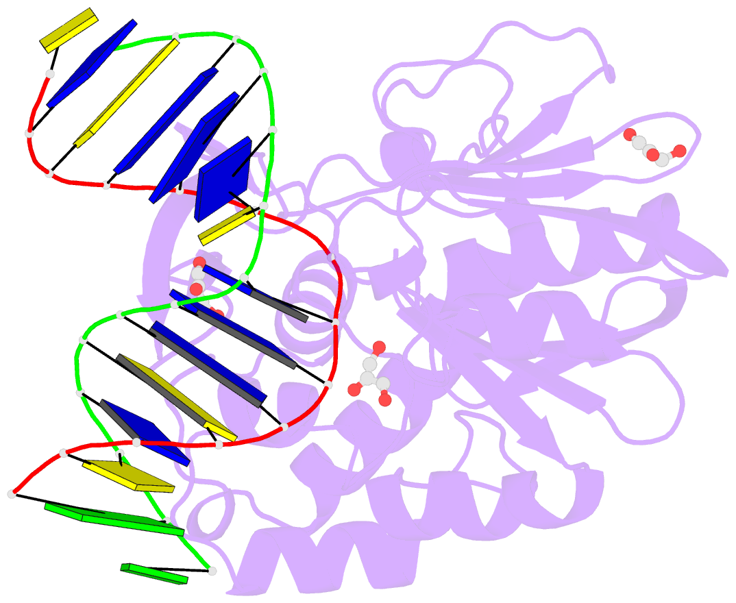
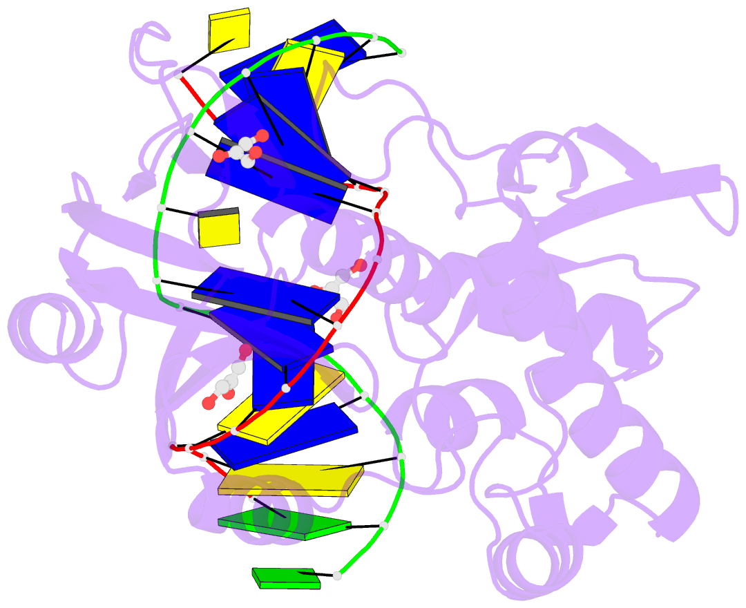
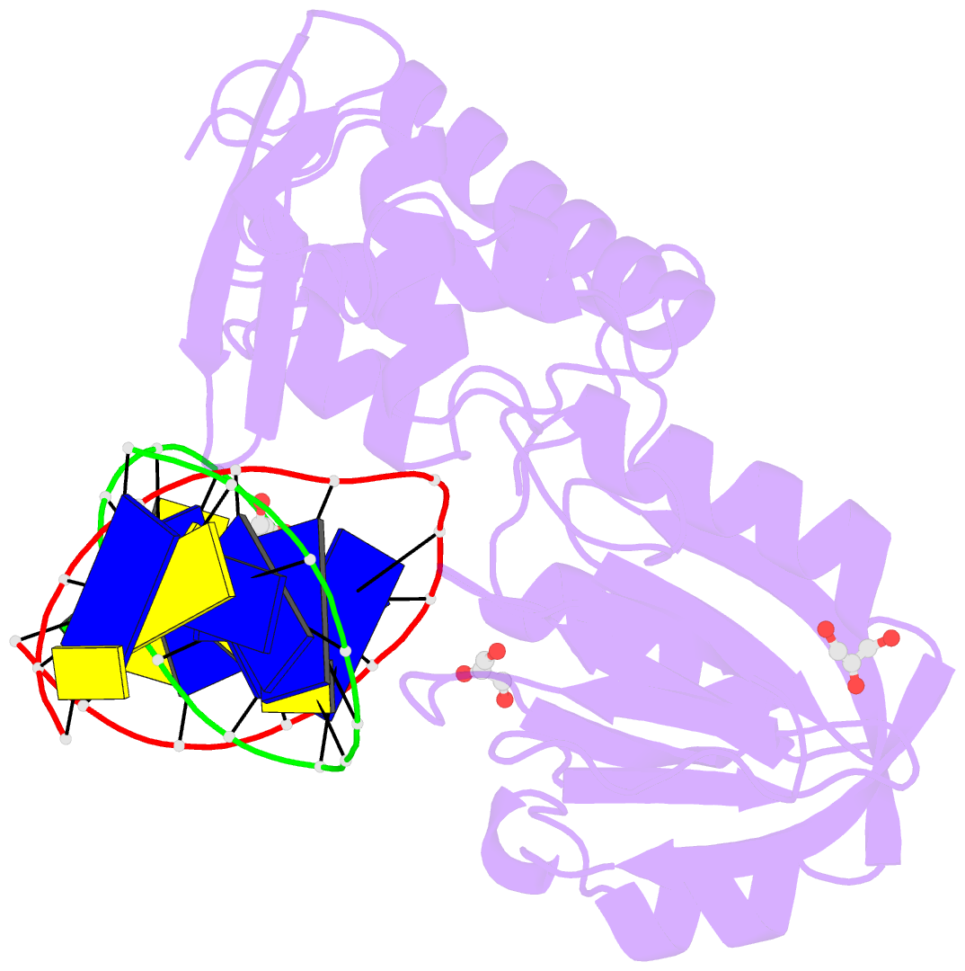
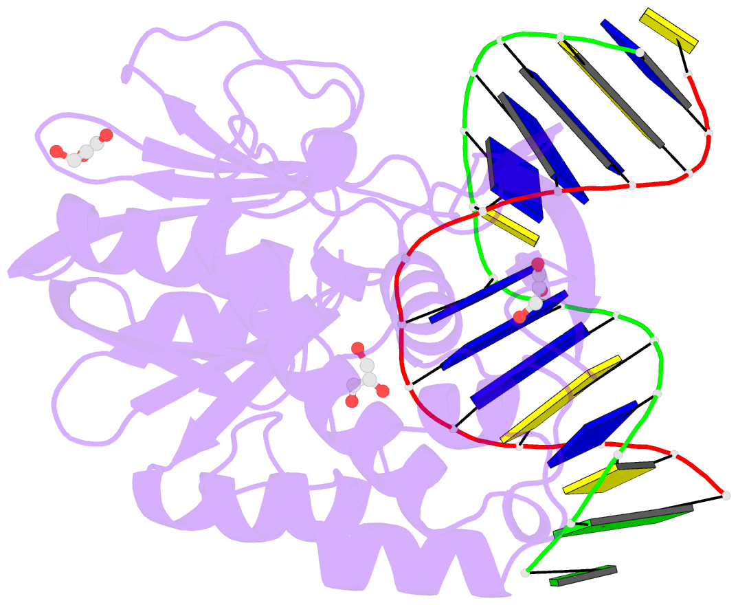
Summary information and primary citation
- PDB-id
- 1pm5; SNAP-derived features in text and JSON formats;
DNAproDB
- Class
- hydrolase-DNA
- Method
- X-ray (1.95 Å)
- Summary
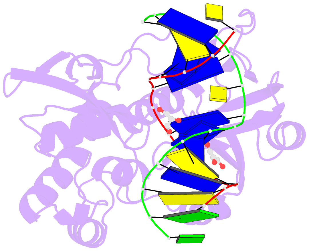
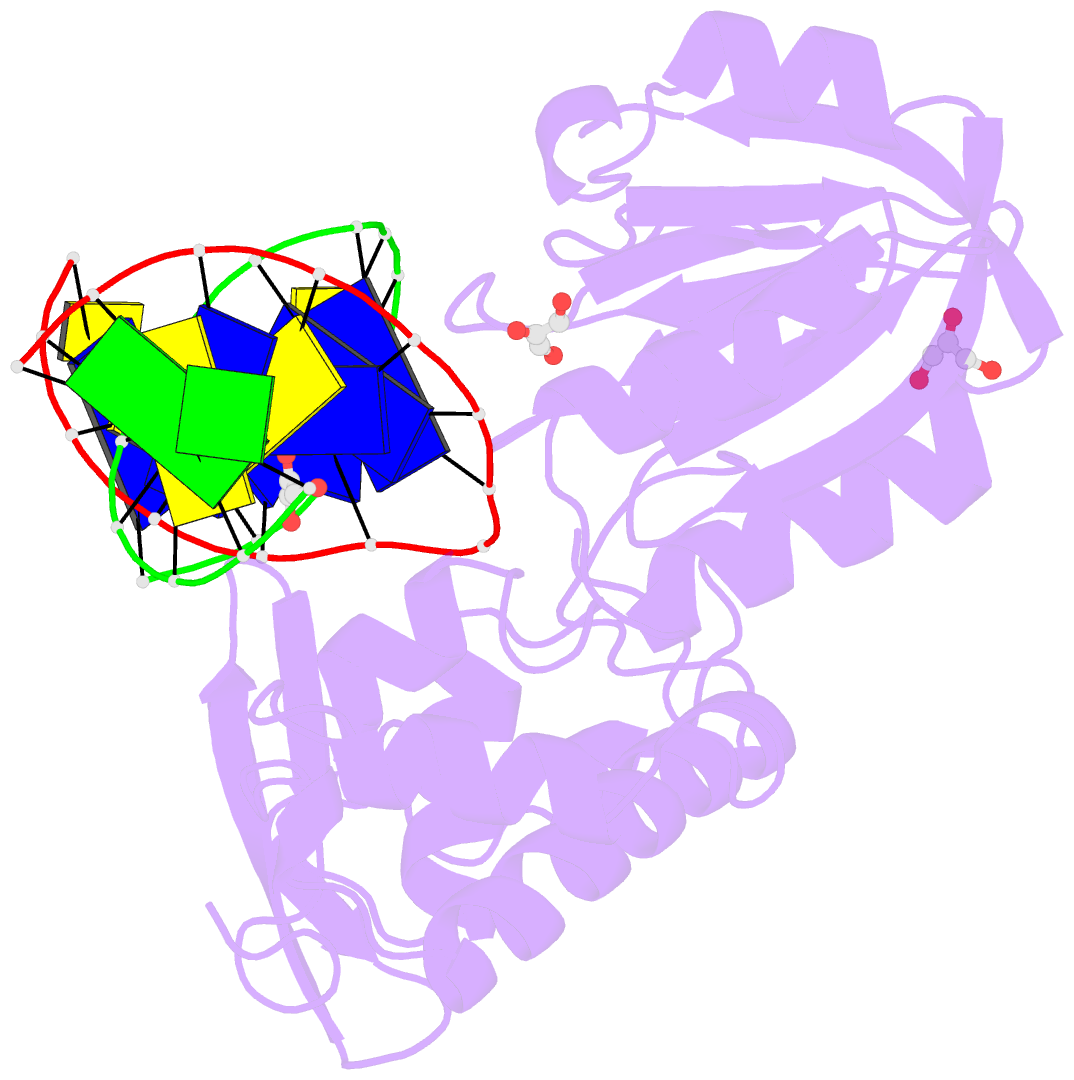
- Crystal structure of wild type lactococcus lactis fpg complexed to a tetrahydrofuran containing DNA
- Reference
- Pereira de Jesus K, Serre L, Zelwer C, Castaing B (2005): "Structural insights into abasic site for Fpg specific binding and catalysis: comparative high-resolution crystallographic studies of Fpg bound to various models of abasic site analogues-containing DNA." Nucleic Acids Res., 33, 5936-5944. doi: 10.1093/nar/gki879.
- Abstract
- Fpg is a DNA glycosylase that recognizes and excises the mutagenic 8-oxoguanine (8-oxoG) and the potentially lethal formamidopyrimidic residues (Fapy). Fpg is also associated with an AP lyase activity which successively cleaves the abasic (AP) site at the 3' and 5' sides by betadelta-elimination. Here, we present the high-resolution crystal structures of the wild-type and the P1G defective mutant of Fpg from Lactococcus lactis bound to 14mer DNA duplexes containing either a tetrahydrofuran (THF) or 1,3-propanediol (Pr) AP site analogues. Structures show that THF is less extrahelical than Pr and its backbone C5'-C4'-C3' diverges significantly from those of Pr, rAP, 8-oxodG and FapydG. Clearly, the heterocyclic oxygen of THF is pushed back by the carboxylate of the strictly conserved E2 residue. We can propose that the ring-opened form of the damaged deoxyribose is the structure active form of the sugar for Fpg catalysis process. Both structural and functional data suggest that the first step of catalysis mediated by Fpg involves the expulsion of the O4' leaving group facilitated by general acid catalysis (involving E2), rather than the immediate cleavage of the N-glycosic bond of the damaged nucleoside.