Summary information and primary citation
- PDB-id
- 1q3f; SNAP-derived features in text and JSON formats;
DNAproDB
- Class
- hydrolase-DNA
- Method
- X-ray (1.9 Å)
- Summary
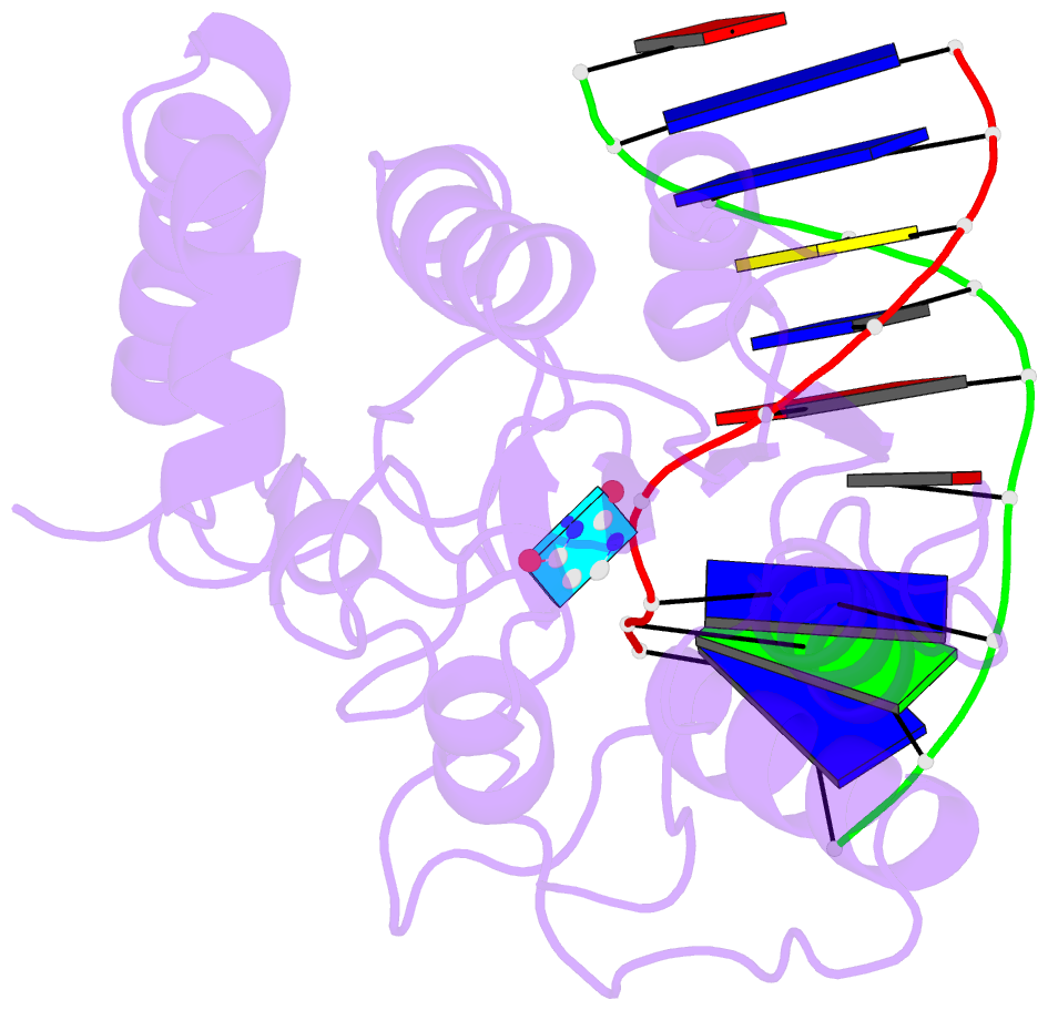
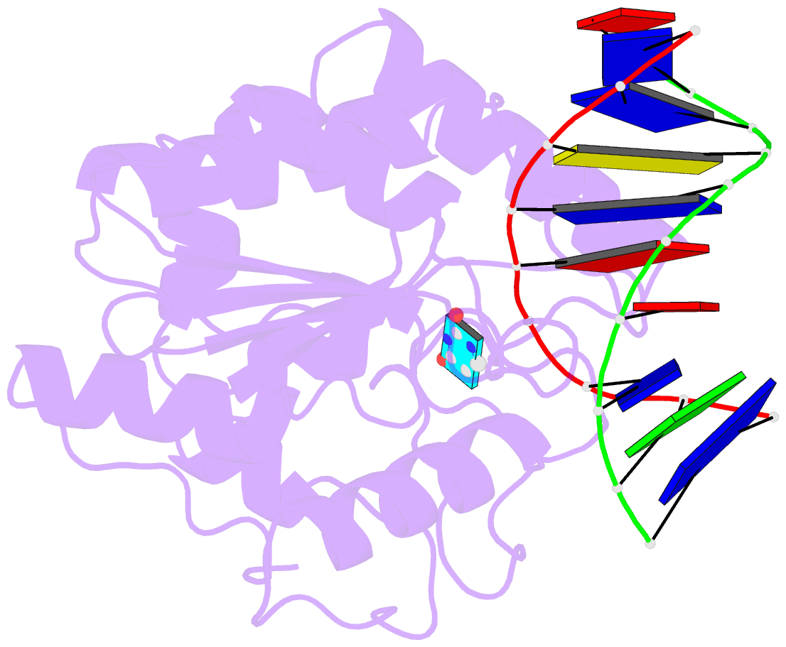
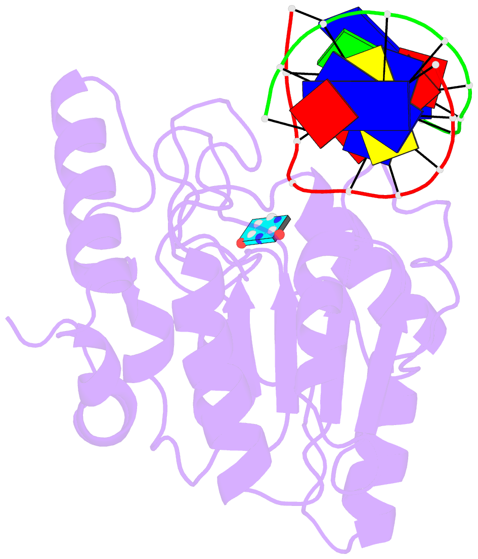
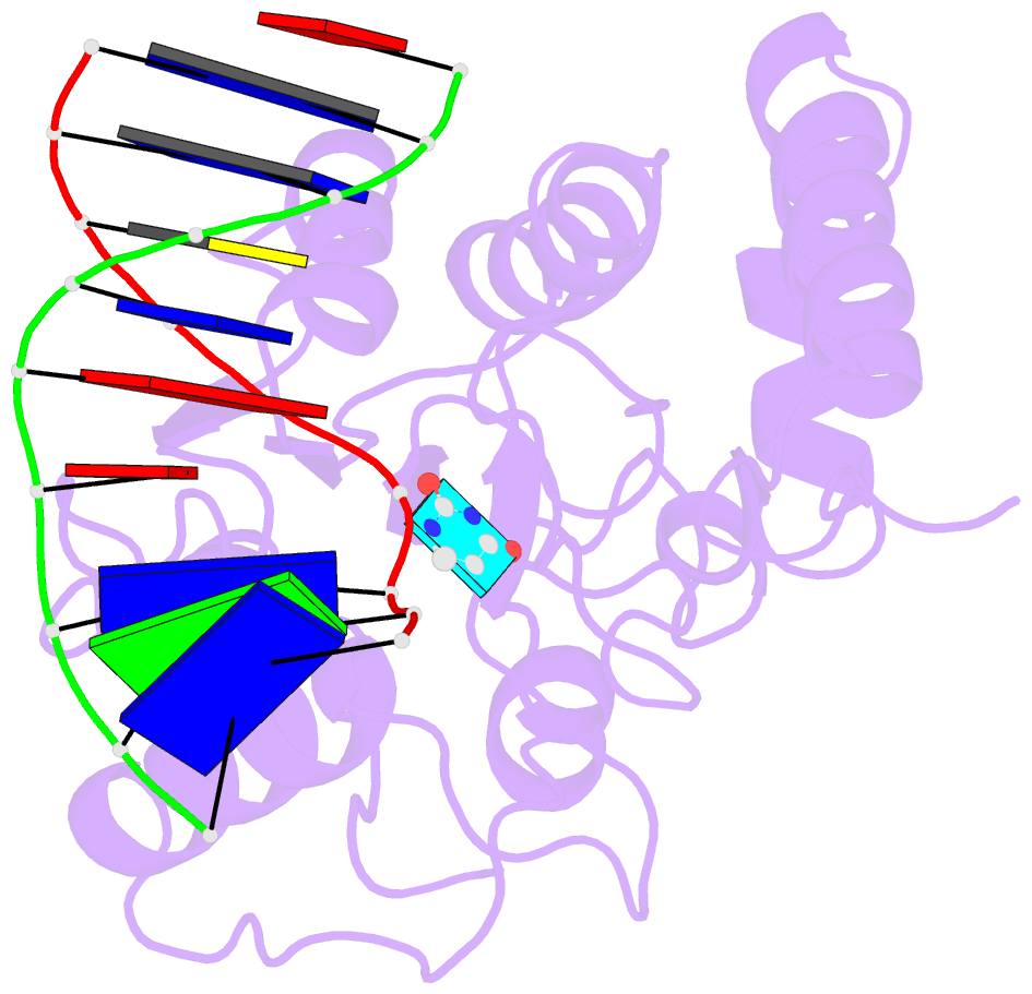
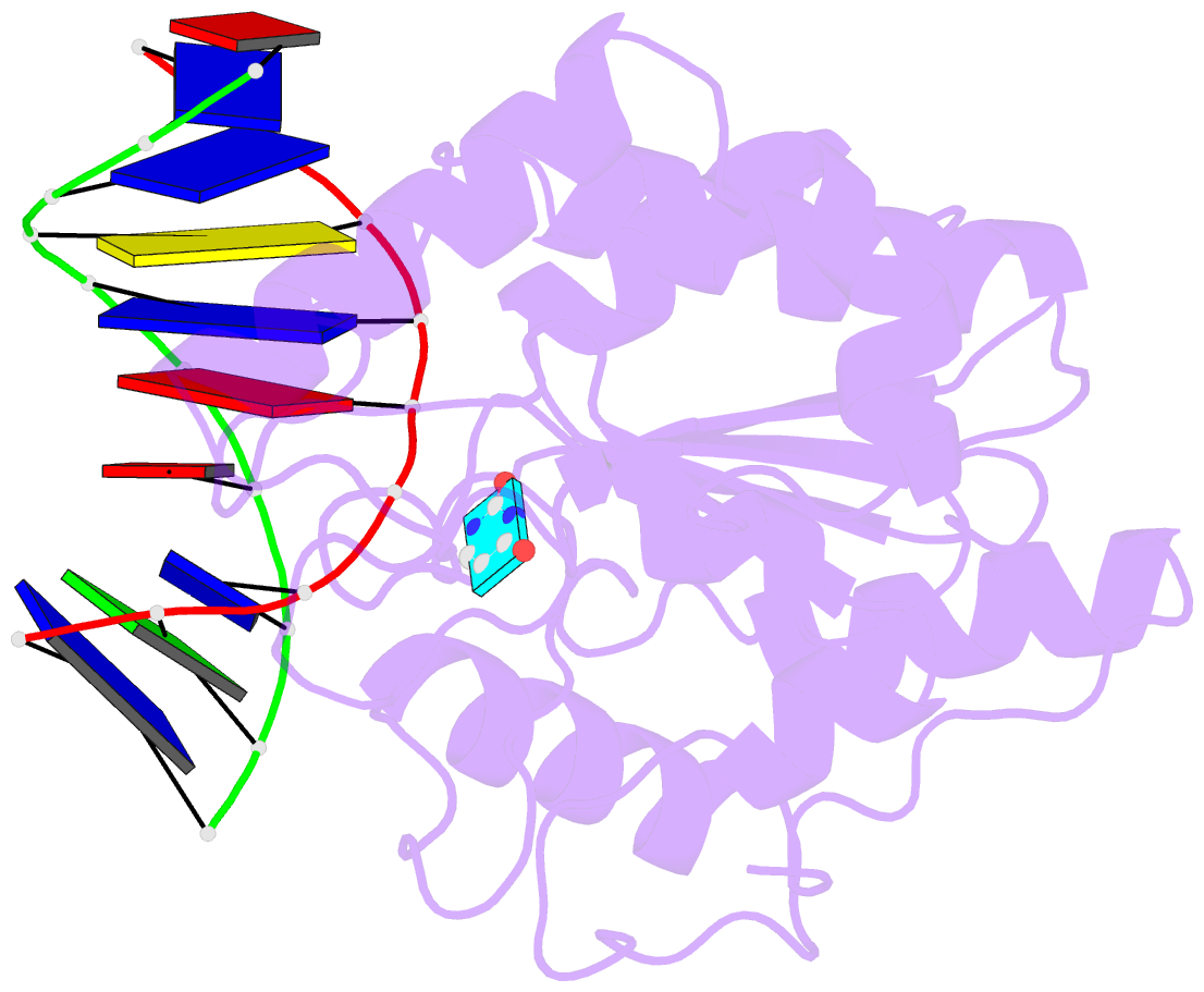
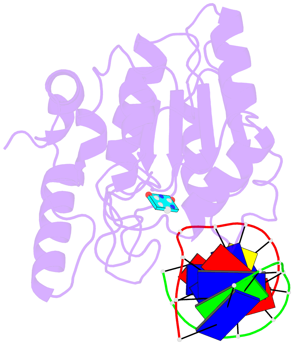
- Uracil DNA glycosylase bound to a cationic 1-aza-2'-deoxyribose-containing DNA
- Reference
- Bianchet MA, Seiple LA, Jiang YL, Ichikawa Y, Amzel LM, Stivers JT (2003): "Electrostatic guidance of glycosyl cation migration along the reaction coordinate of uracil DNA glycosylase." Biochemistry, 42, 12455-12460. doi: 10.1021/bi035372+.
- Abstract
- The DNA repair enzyme uracil DNA glycosylase has been crystallized with a cationic 1-aza-2'-deoxyribose-containing DNA that mimics the ultimate transition state of the reaction in which the water nucleophile attacks the anomeric center of the oxacarbenium ion-uracil anion reaction intermediate. Comparison with substrate and product structures, and the previous structure of the intermediate determined by kinetic isotope effects, reveals an exquisite example of geometric strain, least atomic motion, and electrophile migration in biological catalysis. This structure provides a rare opportunity to reconstruct the detailed structural transformations that occur along an enzymatic reaction coordinate.