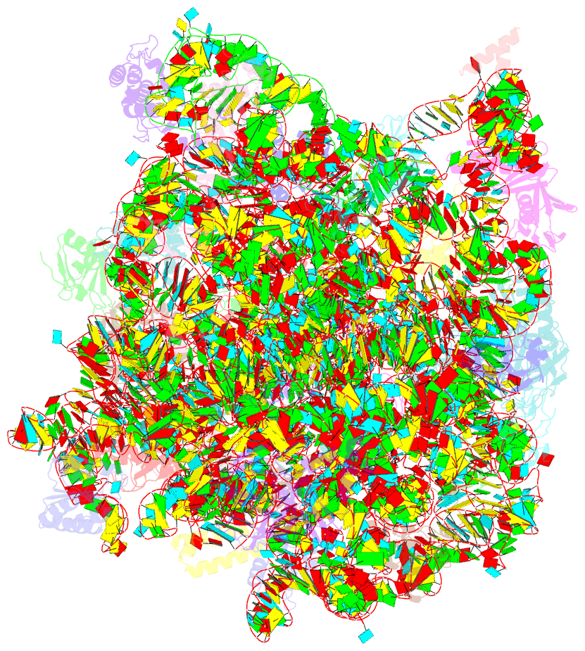
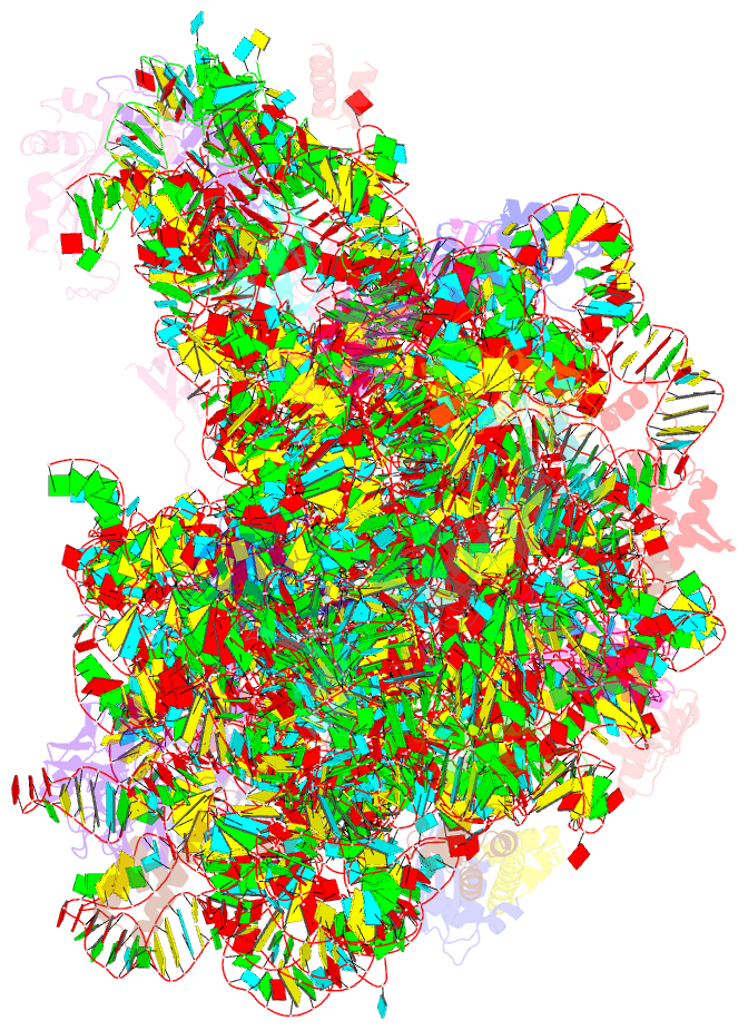
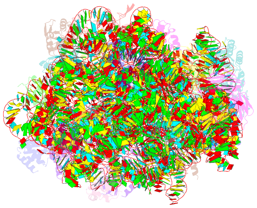
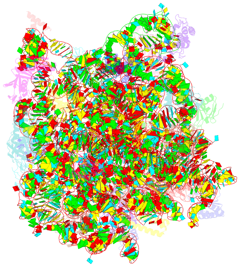
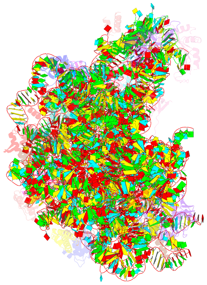
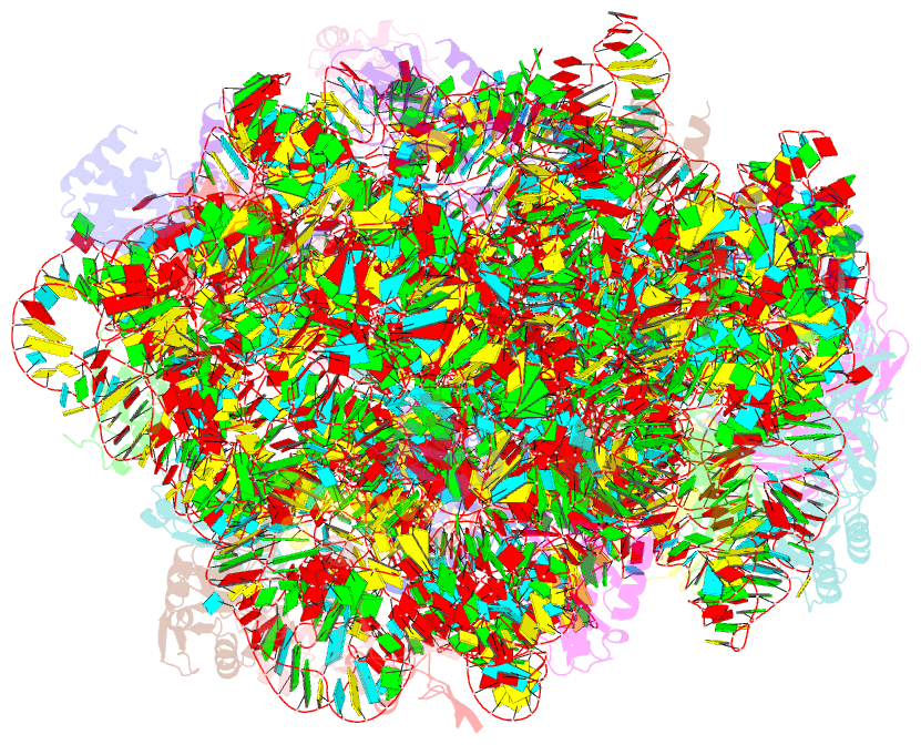
Summary information and primary citation
- PDB-id
- 1q7y; SNAP-derived features in text and JSON formats;
DNAproDB
- Class
- ribosome
- Method
- X-ray (3.2 Å)
- Summary
- Crystal structure of ccdap-puromycin bound at the peptidyl transferase center of the 50s ribosomal subunit
- Reference
- Hansen JL, Schmeing TM, Moore PB, Steitz TA (2002): "Structural Insights Into Peptide Bond Formation." Proc.Natl.Acad.Sci.USA, 99, 11670-11675. doi: 10.1073/pnas.172404099.
- Abstract
- The large ribosomal subunit catalyzes peptide bond formation and will do so by using small aminoacyl- and peptidyl-RNA fragments of tRNA. We have refined at 3-A resolution the structures of both A and P site substrate and product analogues, as well as an intermediate analogue, bound to the Haloarcula marismortui 50S ribosomal subunit. A P site substrate, CCA-Phe-caproic acid-biotin, binds equally to both sites, but in the presence of sparsomycin binds only to the P site. The CCA portions of these analogues are bound identically by either the A or P loop of the 23S rRNA. Combining the separate P and A site substrate complexes into one model reveals interactions that may occur when both are present simultaneously. The alpha-NH(2) group of an aminoacylated fragment in the A site forms one hydrogen bond with the N3 of A2486 (2451) and may form a second hydrogen bond either with the 2' OH of the A-76 ribose in the P site or with the 2' OH of A2486 (2451). These interactions position the alpha amino group adjacent to the carbonyl carbon of esterified P site substrate in an orientation suitable for a nucleophilic attack.