Summary information and primary citation
- PDB-id
- 1qa6; SNAP-derived features in text and JSON formats;
DNAproDB
- Class
- ribosome
- Method
- X-ray (2.8 Å)
- Summary
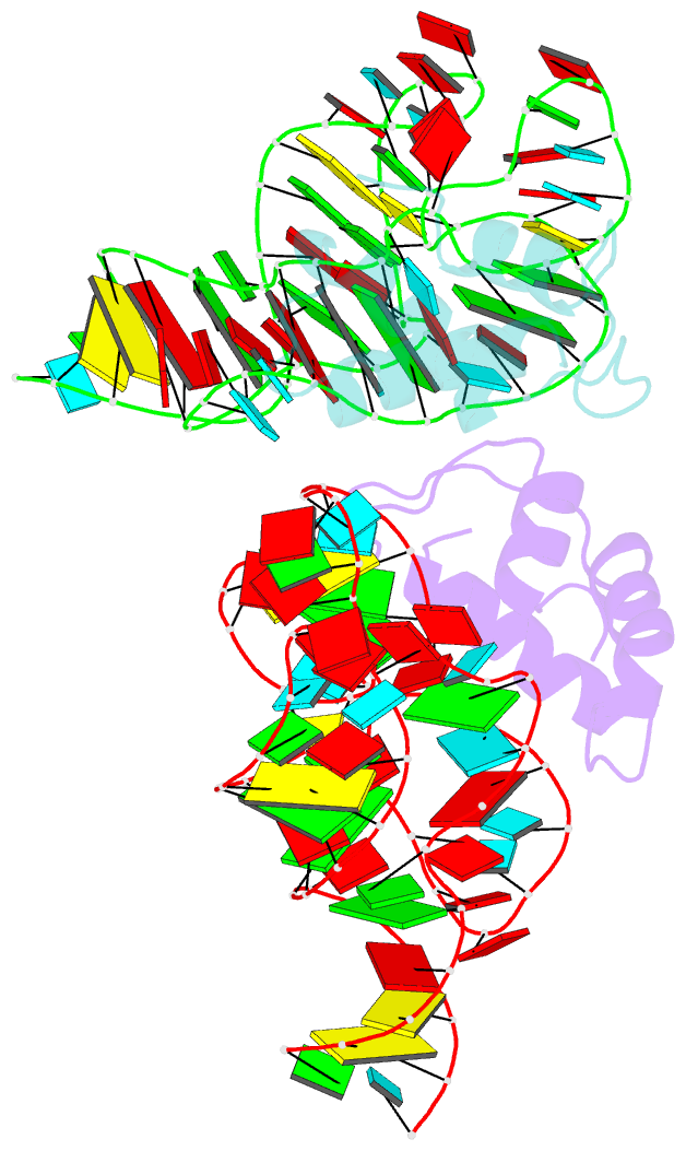
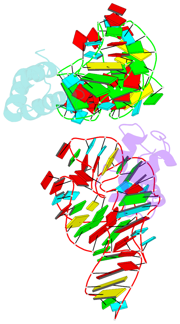
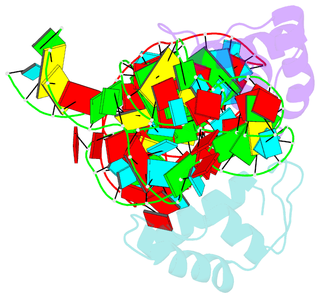
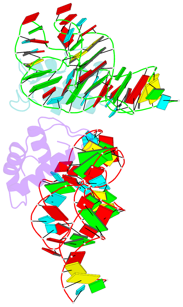
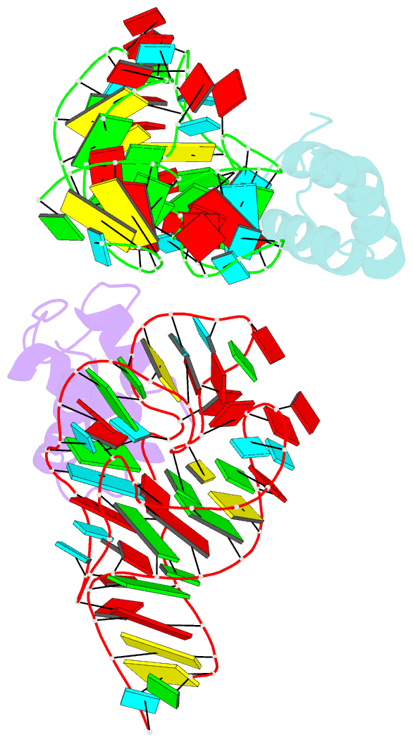
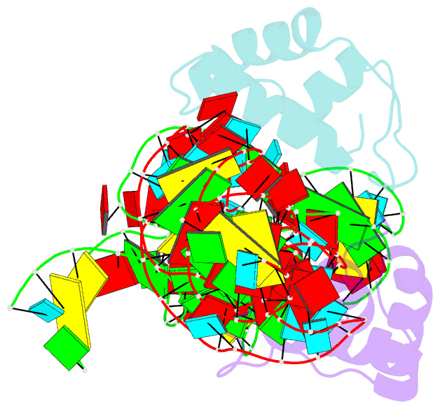
- Crystal structure of a conserved ribosomal protein-RNA complex
- Reference
- Conn GL, Draper DE, Lattman EE, Gittis AG (1999): "Crystal structure of a conserved ribosomal protein-RNA complex." Science, 284, 1171-1174. doi: 10.1126/science.284.5417.1171.
- Abstract
- The structure of a highly conserved complex between a 58-nucleotide domain of large subunit ribosomal RNA and the RNA-binding domain of ribosomal protein L11 has been solved at 2.8 angstrom resolution. It reveals a precisely folded RNA structure that is stabilized by extensive tertiary contacts and contains an unusually large core of stacked bases. A bulge loop base from one hairpin of the RNA is intercalated into the distorted major groove of another helix; the protein locks this tertiary interaction into place by binding to the intercalated base from the minor groove side. This direct interaction with a key ribosomal RNA tertiary interaction suggests that part of the role of L11 is to stabilize an unusual RNA fold within the ribosome.