Summary information and primary citation
- PDB-id
- 1qbj; SNAP-derived features in text and JSON formats;
DNAproDB
- Class
- hydrolase-DNA
- Method
- X-ray (2.1 Å)
- Summary
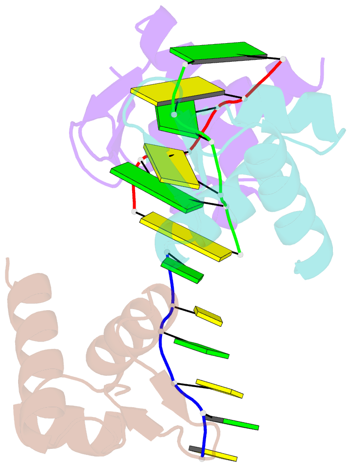
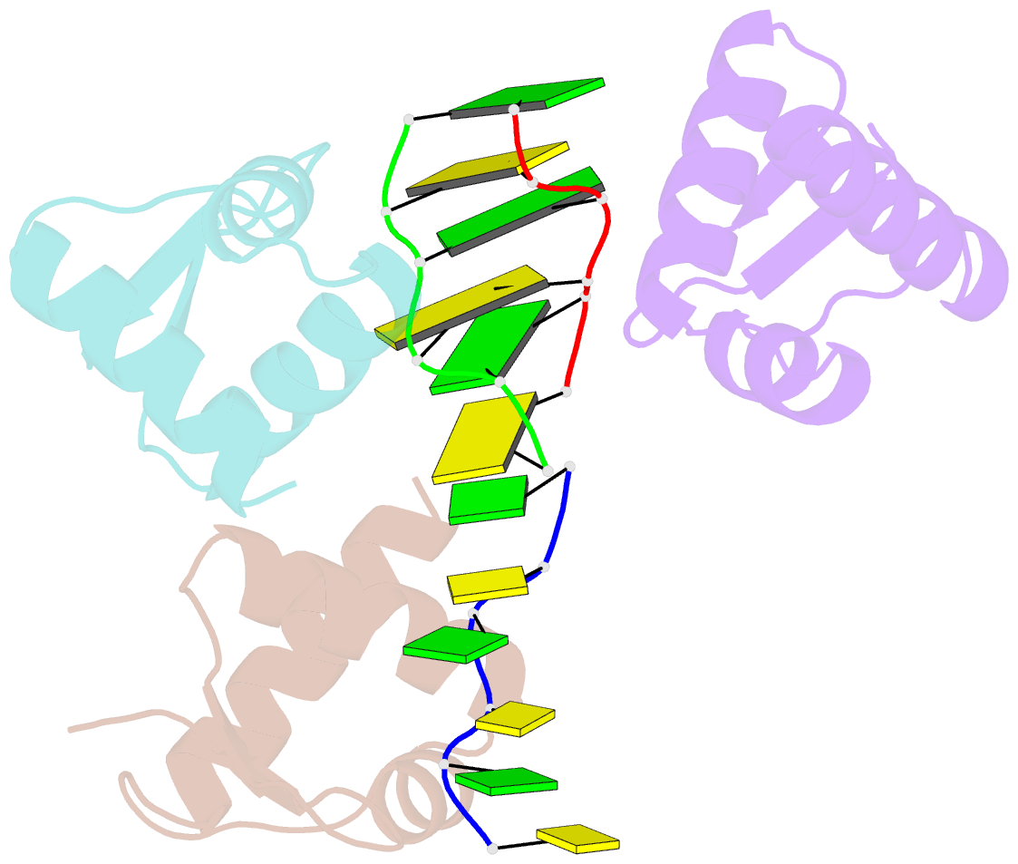
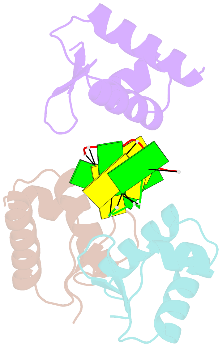
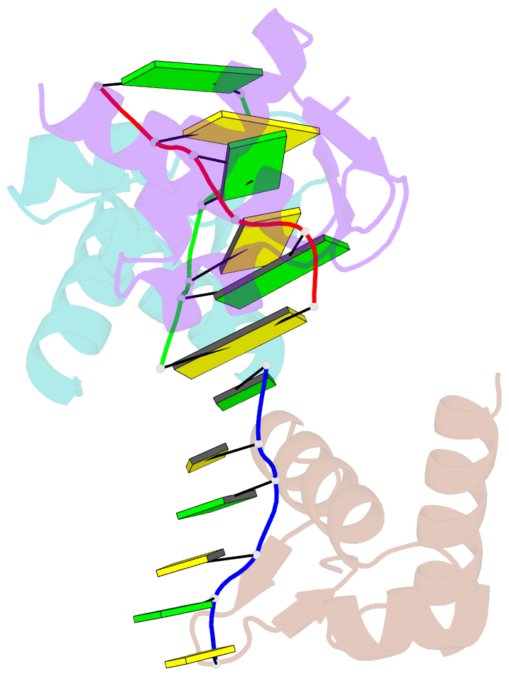
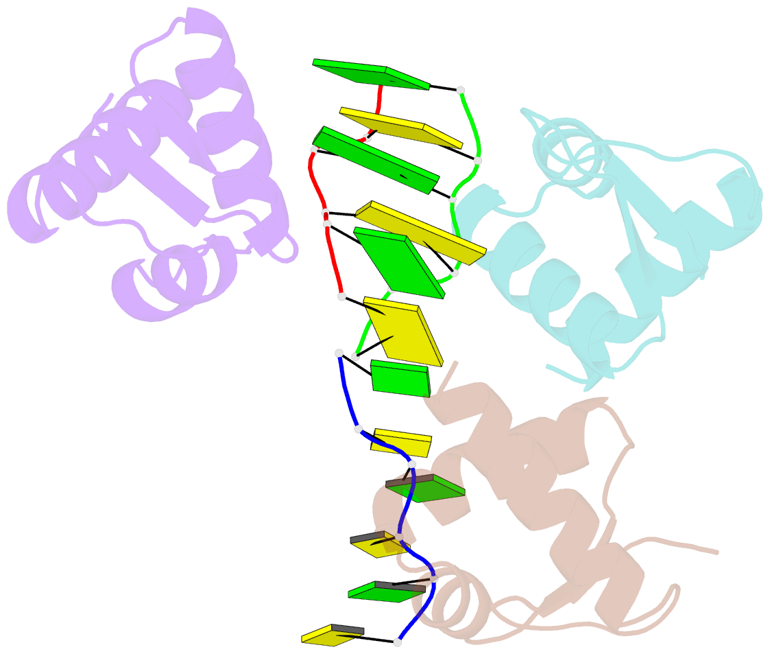
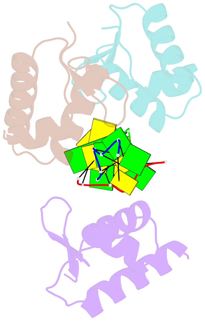
- Crystal structure of the zalpha z-DNA complex
- Reference
- Schwartz T, Rould MA, Lowenhaupt K, Herbert A, Rich A (1999): "Crystal structure of the Zalpha domain of the human editing enzyme ADAR1 bound to left-handed Z-DNA." Science, 284, 1841-1845. doi: 10.1126/science.284.5421.1841.
- Abstract
- The editing enzyme double-stranded RNA adenosine deaminase includes a DNA binding domain, Zalpha, which is specific for left-handed Z-DNA. The 2.1 angstrom crystal structure of Zalpha complexed to DNA reveals that the substrate is in the left-handed Z conformation. The contacts between Zalpha and Z-DNA are made primarily with the "zigzag" sugar-phosphate backbone, which provides a basis for the specificity for the Z conformation. A single base contact is observed to guanine in the syn conformation, characteristic of Z-DNA. Intriguingly, the helix-turn-helix motif, frequently used to recognize B-DNA, is used by Zalpha to contact Z-DNA.