Summary information and primary citation
- PDB-id
- 1rbj; SNAP-derived features in text and JSON formats;
DNAproDB
- Class
- hydrolase-DNA
- Method
- X-ray (2.7 Å)
- Summary
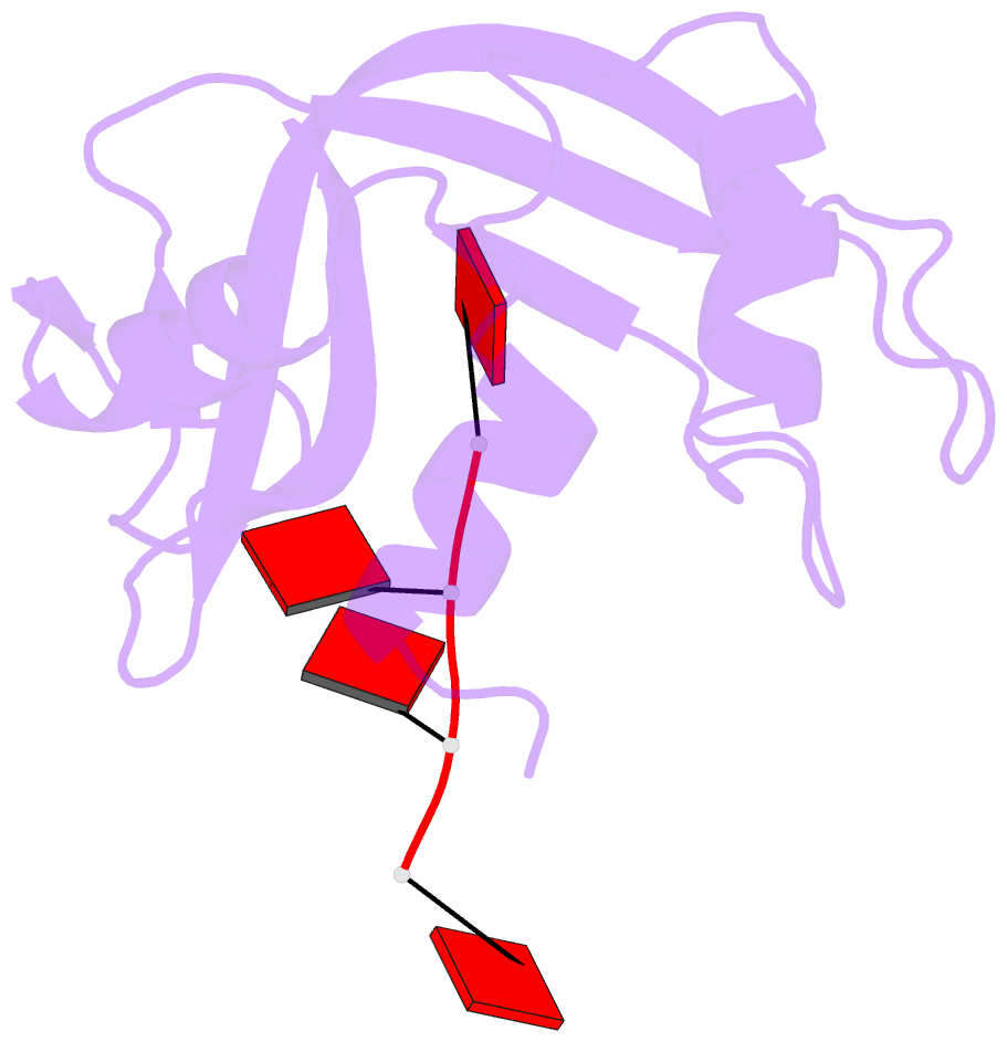
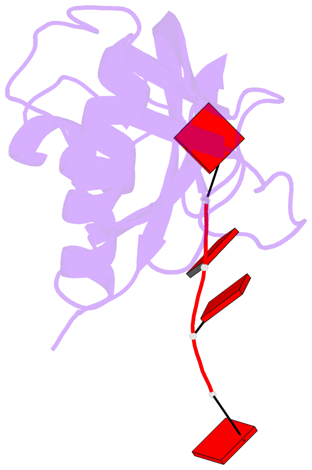
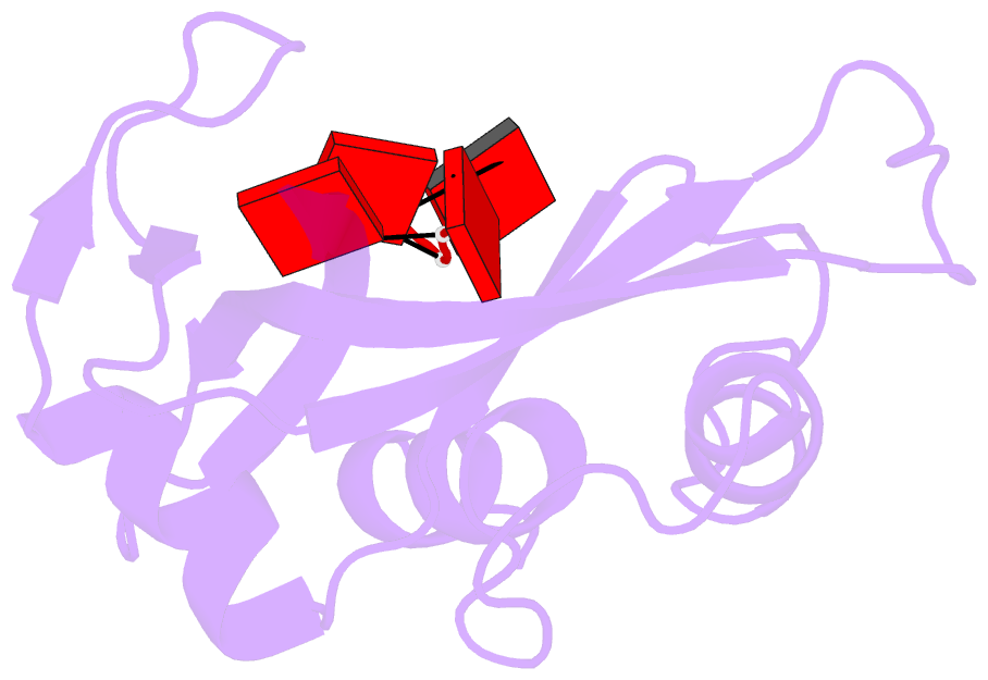
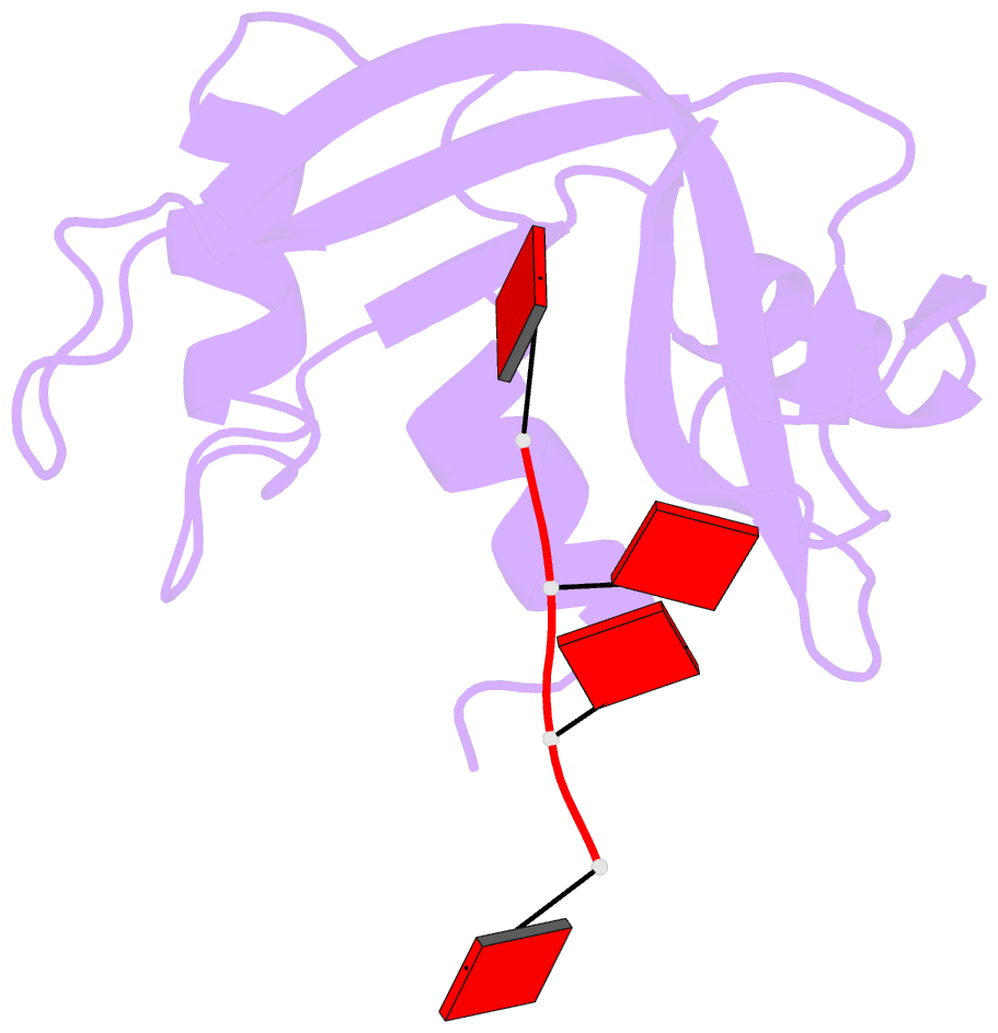
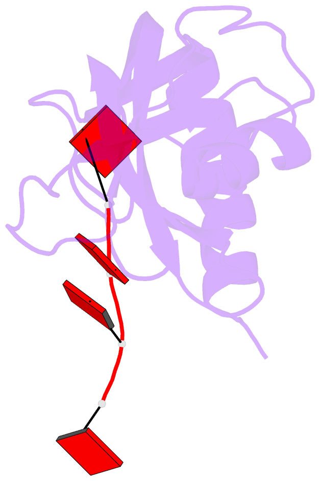
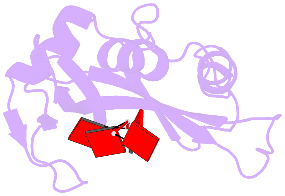
- Ribonuclease b complex with d(tetra-(deoxy-adenylate))
- Reference
- Ko TP, Williams R, McPherson A (1996): "Structure of a ribonuclease B+d(pA)4 complex." Acta Crystallogr.,Sect.D, 52, 160-164. doi: 10.1107/S0907444995009127.
- Abstract
- The structure of a tetragonal crystal of bovine pancreatic RNase B complexed with d(pA)(4) was determined by molecular replacement and difference Fourier methods. This crystal belongs to space group P4(1)2(1)2 and has unit-cell dimensions a = b = 44.5, c = 156.5 A. The model consists of the enzyme and a tetranucleotide with fractional occupancies, suggesting multiple modes of oligonucleotide binding. It does not include any polysaccharide residues or solvent molecules. After refinement at 2.7 A, the R value was 0.163 with acceptable stereochemistry. The model illustrates a set of well defined interactions for substrate binding, particularly between the central dinucleotide and the enzyme.