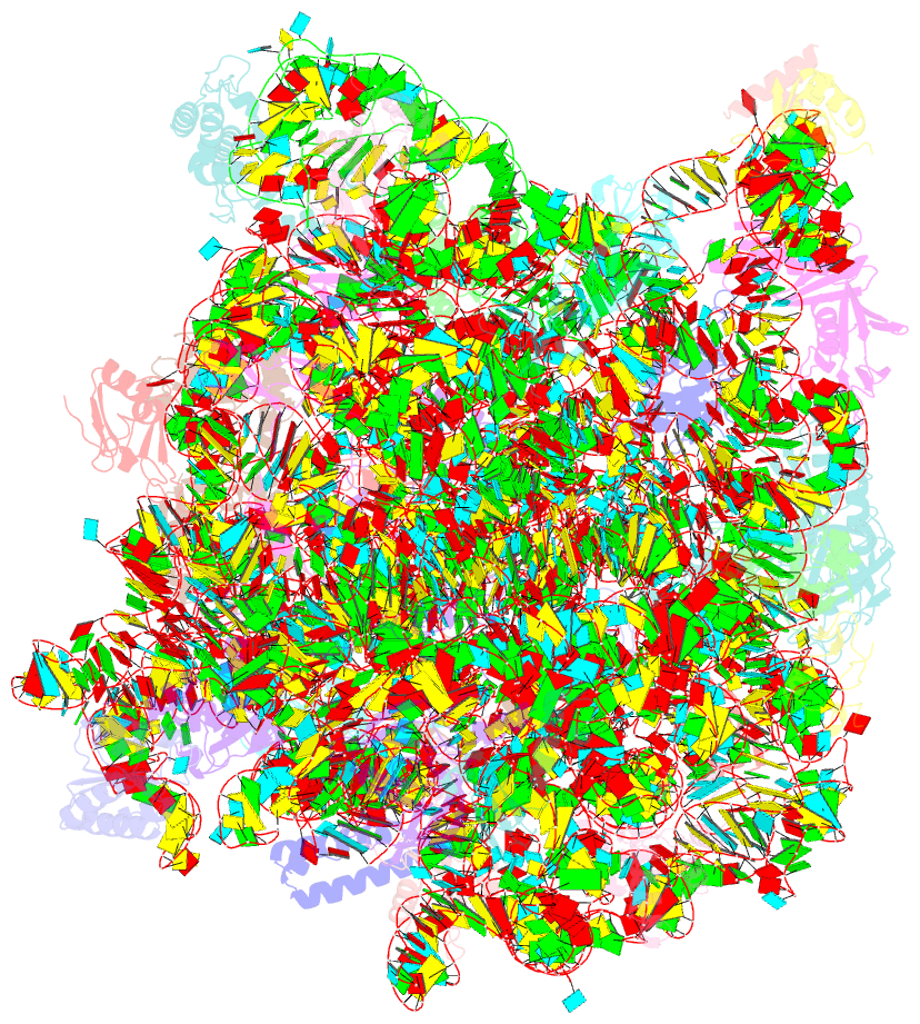
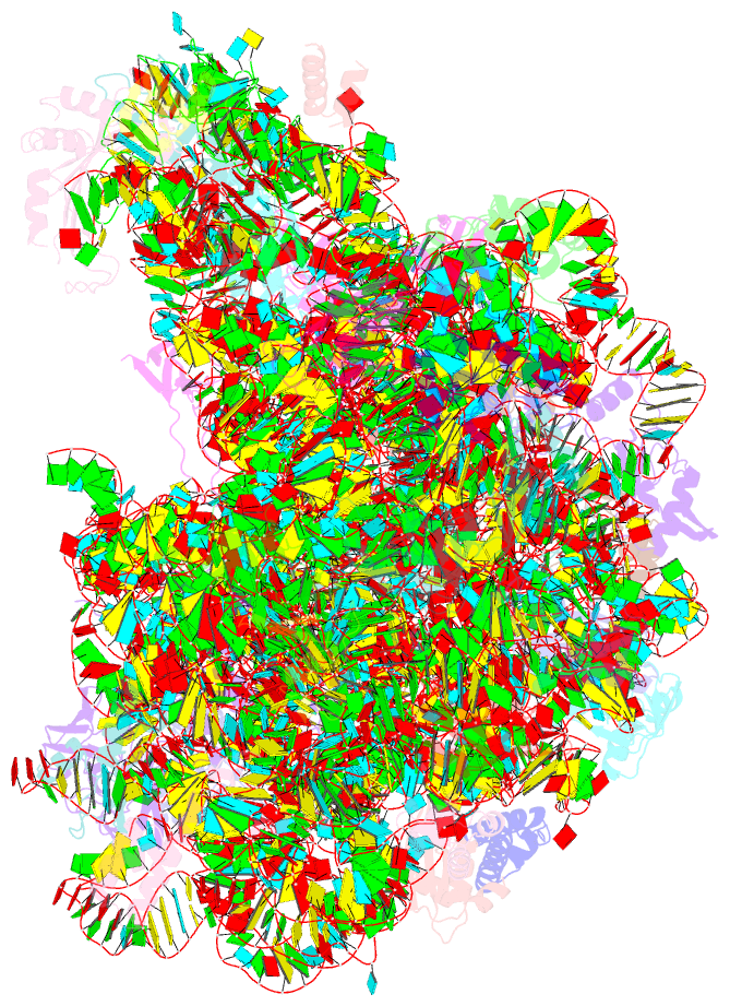
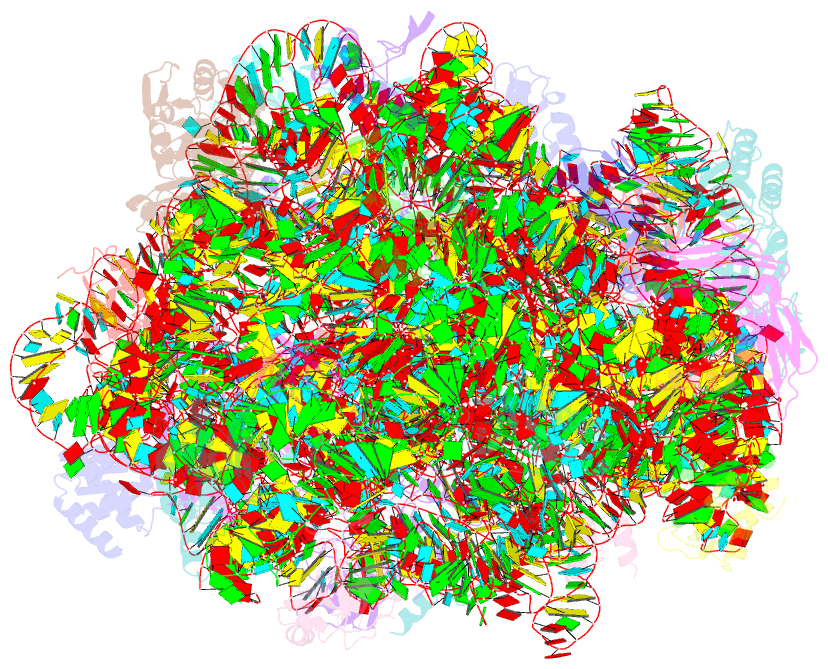
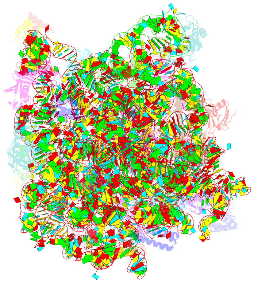
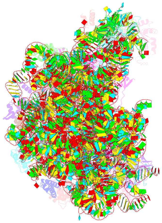
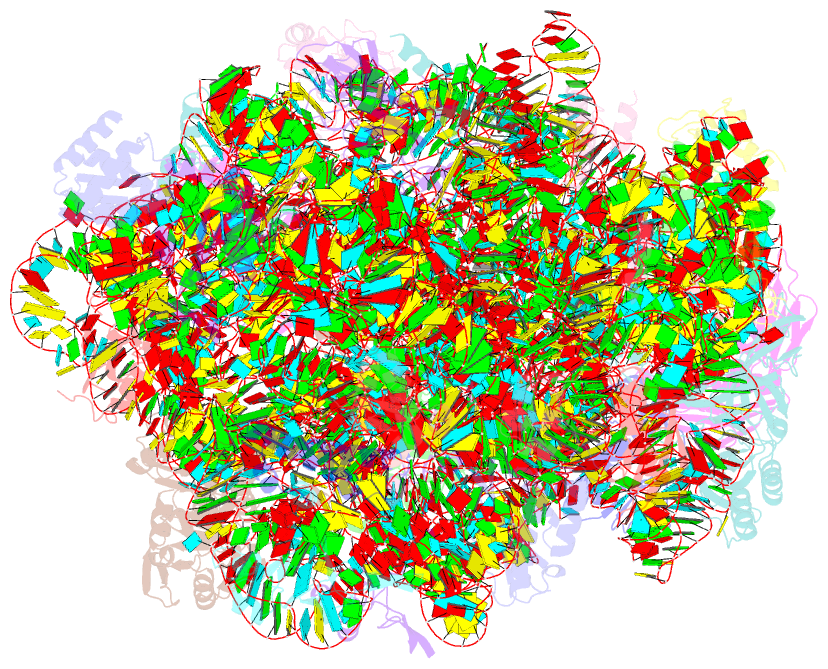
Summary information and primary citation
- PDB-id
- 1s72; SNAP-derived features in text and JSON formats;
DNAproDB
- Class
- ribosome
- Method
- X-ray (2.4 Å)
- Summary
- Refined crystal structure of the haloarcula marismortui large ribosomal subunit at 2.4 angstrom resolution
- Reference
- Klein DJ, Moore PB, Steitz TA (2004): "The Roles of Ribosomal Proteins in the Structure, Assembly and Evolution of the Large Ribosomal Subunit." J.Mol.Biol., 340, 141-177. doi: 10.1016/j.jmb.2004.03.076.
- Abstract
- The structures of ribosomal proteins and their interactions with RNA have been examined in the refined crystal structure of the Haloarcula marismortui large ribosomal subunit. The protein structures fall into six groups based on their topology. The 50S subunit proteins function primarily to stabilize inter-domain interactions that are necessary to maintain the subunit's structural integrity. An extraordinary variety of protein-RNA interactions is observed. Electrostatic interactions between numerous arginine and lysine residues, particularly those in tail extensions, and the phosphate groups of the RNA backbone mediate many protein-RNA contacts. Base recognition occurs via both the minor groove and widened major groove of RNA helices, as well as through hydrophobic binding pockets that capture bulged nucleotides and through insertion of amino acid residues into hydrophobic crevices in the RNA. Primary binding sites on contiguous RNA are identified for 20 of the 50S ribosomal proteins, which along with few large protein-protein interfaces, suggest the order of assembly for some proteins and that the protein extensions fold cooperatively with RNA. The structure supports the hypothesis of co-transcriptional assembly, centered around L24 in domain I. Finally, comparing the structures and locations of the 50S ribosomal proteins from H.marismortui and D.radiodurans revealed striking examples of molecular mimicry. These comparisons illustrate that identical RNA structures can be stabilized by unrelated proteins.