Summary information and primary citation
- PDB-id
- 1tez; SNAP-derived features in text and JSON formats;
DNAproDB
- Class
- lyase-DNA
- Method
- X-ray (1.8 Å)
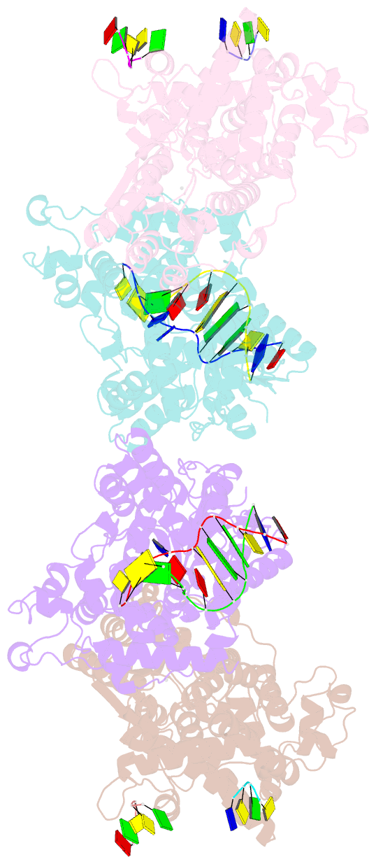
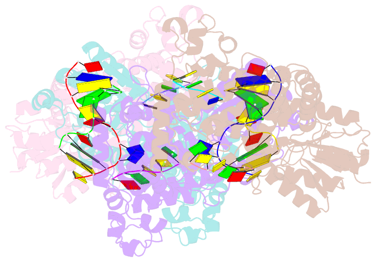
- Summary
- Complex between DNA and the DNA photolyase from anacystis nidulans
- Reference
- Mees A, Klar T, Gnau P, Hennecke U, Eker APM, Carell T, Essen L-O (2004): "Crystal structure of a photolyase bound to a CPD-like DNA lesion after in situ repair." Science, 306, 1789-1793. doi: 10.1126/science.1101598.
- Abstract
- DNA photolyases use light energy to repair DNA that comprises ultraviolet-induced lesions such as the cis-syn cyclobutane pyrimidine dimers (CPDs). Here we report the crystal structure of a DNA photolyase bound to duplex DNA that is bent by 50 degrees and comprises a synthetic CPD lesion. This CPD lesion is flipped into the active site and split there into two thymines by synchrotron radiation at 100 K. Although photolyases catalyze blue light-driven CPD cleavage only above 200 K, this structure apparently mimics a structural substate during light-driven DNA repair in which back-flipping of the thymines into duplex DNA has not yet taken place.