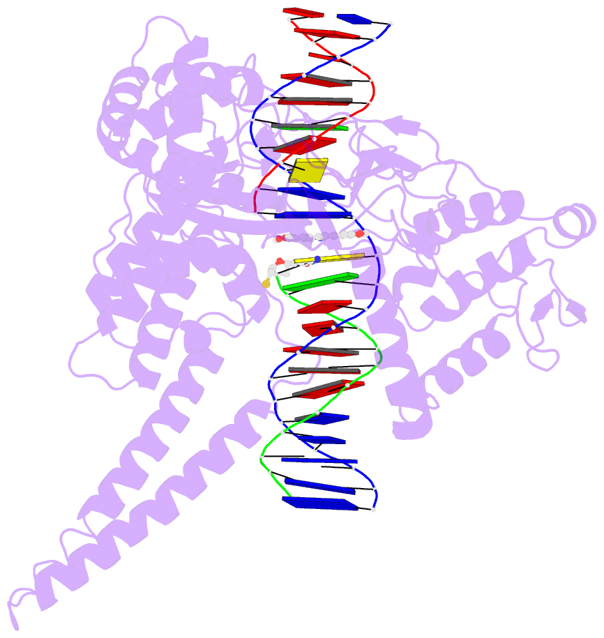
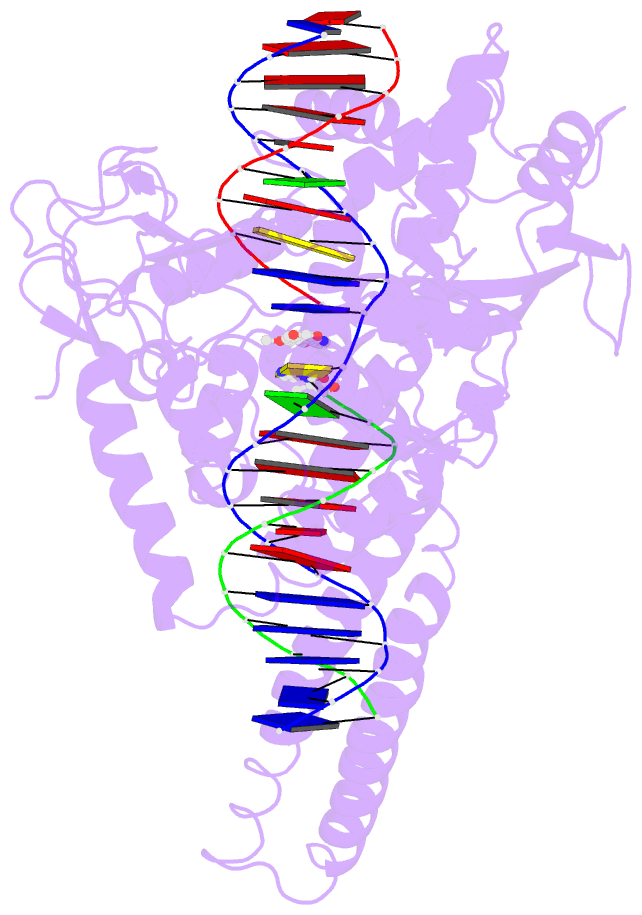
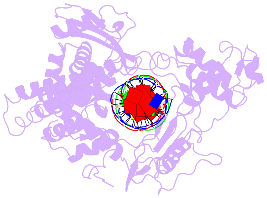
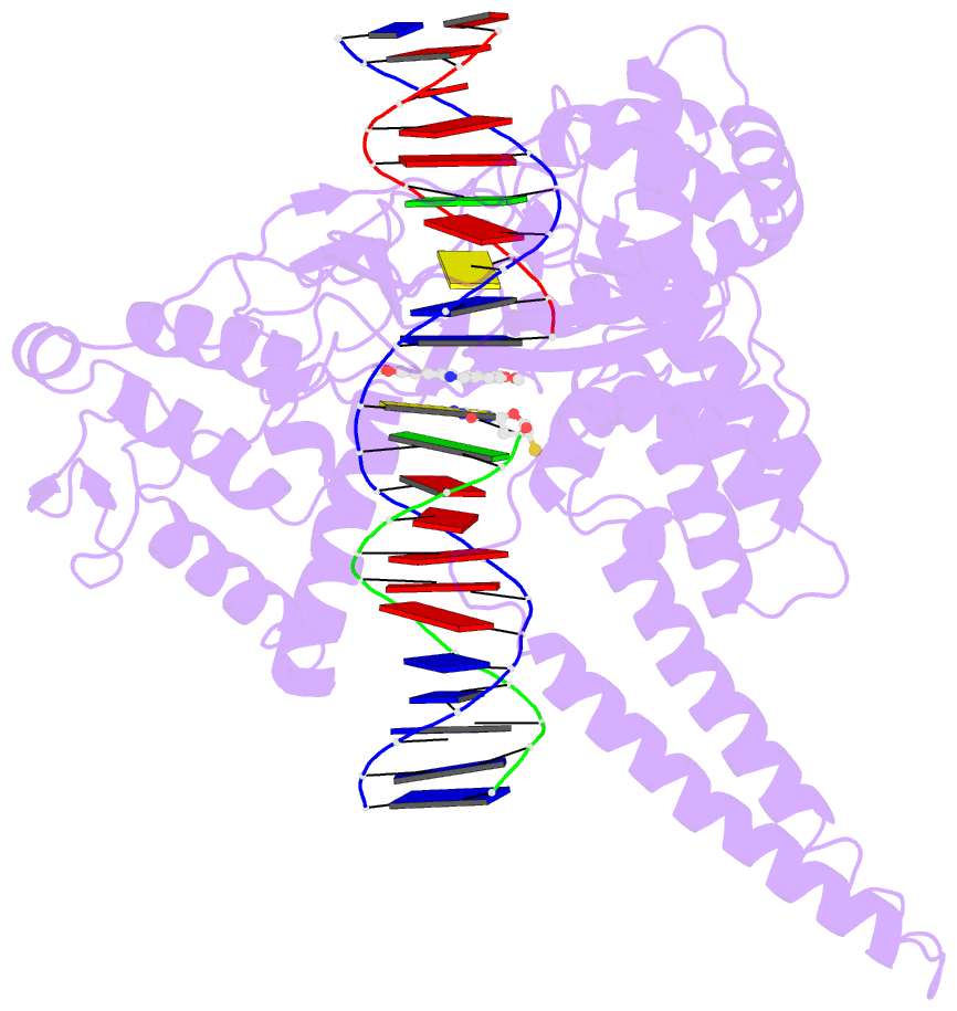
Summary information and primary citation
- PDB-id
- 1tl8; SNAP-derived features in text and JSON formats;
DNAproDB
- Class
- isomerase-DNA
- Method
- X-ray (3.1 Å)
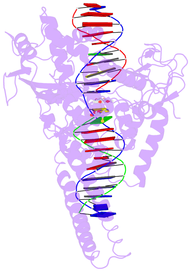
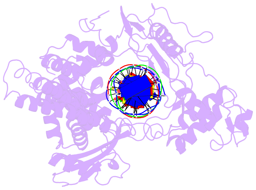
- Summary
- Human DNA topoisomerase i (70 kda) in complex with the indenoisoquinoline ai-iii-52 and covalent complex with a 22 base pair DNA duplex
- Reference
- Ioanoviciu A, Antony S, Pommier Y, Staker BL, Stewart L, Cushman M (2005): "Synthesis and Mechanism of Action Studies of a Series of Norindenoisoquinoline Topoisomerase I Poisons Reveal an Inhibitor with a Flipped Orientation in the Ternary DNA-Enzyme-Inhibitor Complex As Determined by X-ray Crystallographic Analysis." J.Med.Chem., 48, 4803-4814. doi: 10.1021/jm050076b.
- Abstract
- Several norindenoisoquinolines substituted with methoxy or methylenedioxy groups have been prepared and their anticancer properties evaluated in cancer cell cultures and in topoisomerase I inhibition assays. 2,3-Dimethoxy-8,9-methylenedioxy-11H-indeno[1,2-c]isoquinoline hydrochloride (14) is a strong topoisomerase I inhibitor and also displays very high cytotoxicity in the NCI cancer cell culture screen (mean graph midpoint of 50 nM). The X-ray crystal structure of norindenoisoquinoline 14 in complex with topoisomerase I and DNA has been solved, providing insight into the structure-activity relationships within this class of new anticancer agents. The number and position of the norindenoisoquinoline substituents have a significant influence on biological activity and demonstrate that substitution on the nitrogen atom is not an absolute requirement for the antitumor effect of the indenoisoquinolines. Removal of the 11-keto group from the lead compound 1 and replacement of the N-alkyllactam with an unsubstituted pyridine ring causes the indenoisoquinoline ring system to flip over in the DNA-enzyme-inhibitor ternary complex. This allows the nitrogen atom to assume the hydrogen bond acceptor role of the 11-keto group, resulting in hydrogen bonding to Arg364.