Summary information and primary citation
- PDB-id
- 1u6b; SNAP-derived features in text and JSON formats;
DNAproDB
- Class
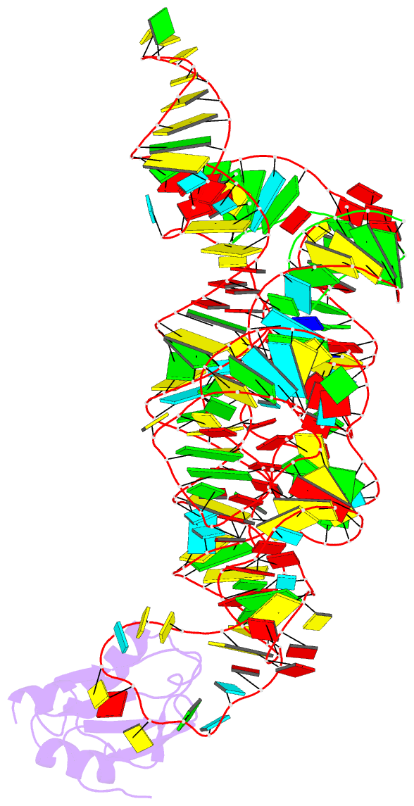
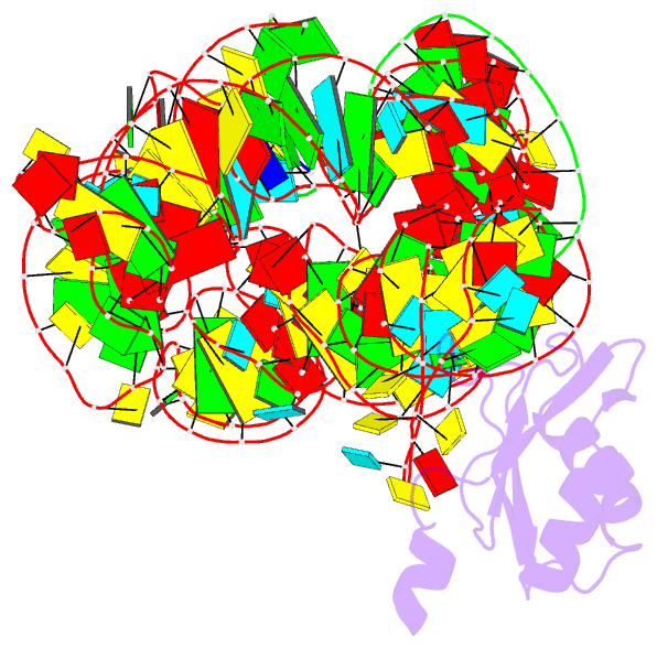
- structural protein-RNA
- Method
- X-ray (3.1 Å)
- Summary
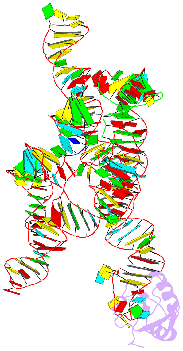
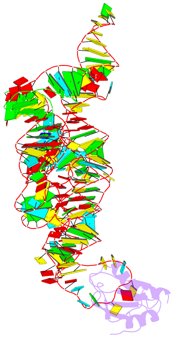
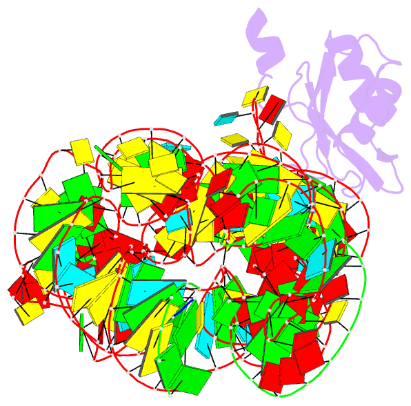
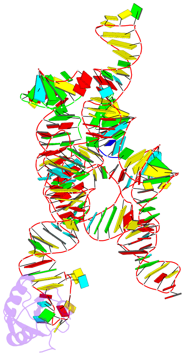
- Crystal structure of a self-splicing group i intron with both exons
- Reference
- Adams PL, Stahley MR, Kosek AB, Wang J, Strobel SA (2004): "Crystal Structure of a Self-Splicing Group I Intron with Both Exons." Nature, 430, 45-50. doi: 10.1038/nature02642.
- Abstract
- The discovery of the RNA self-splicing group I intron provided the first demonstration that not all enzymes are proteins. Here we report the X-ray crystal structure (3.1-A resolution) of a complete group I bacterial intron in complex with both the 5'- and the 3'-exons. This complex corresponds to the splicing intermediate before the exon ligation step. It reveals how the intron uses structurally unprecedented RNA motifs to select the 5'- and 3'-splice sites. The 5'-exon's 3'-OH is positioned for inline nucleophilic attack on the conformationally constrained scissile phosphate at the intron-3'-exon junction. Six phosphates from three disparate RNA strands converge to coordinate two metal ions that are asymmetrically positioned on opposing sides of the reactive phosphate. This structure represents the first splicing complex to include a complete intron, both exons and an organized active site occupied with metal ions.