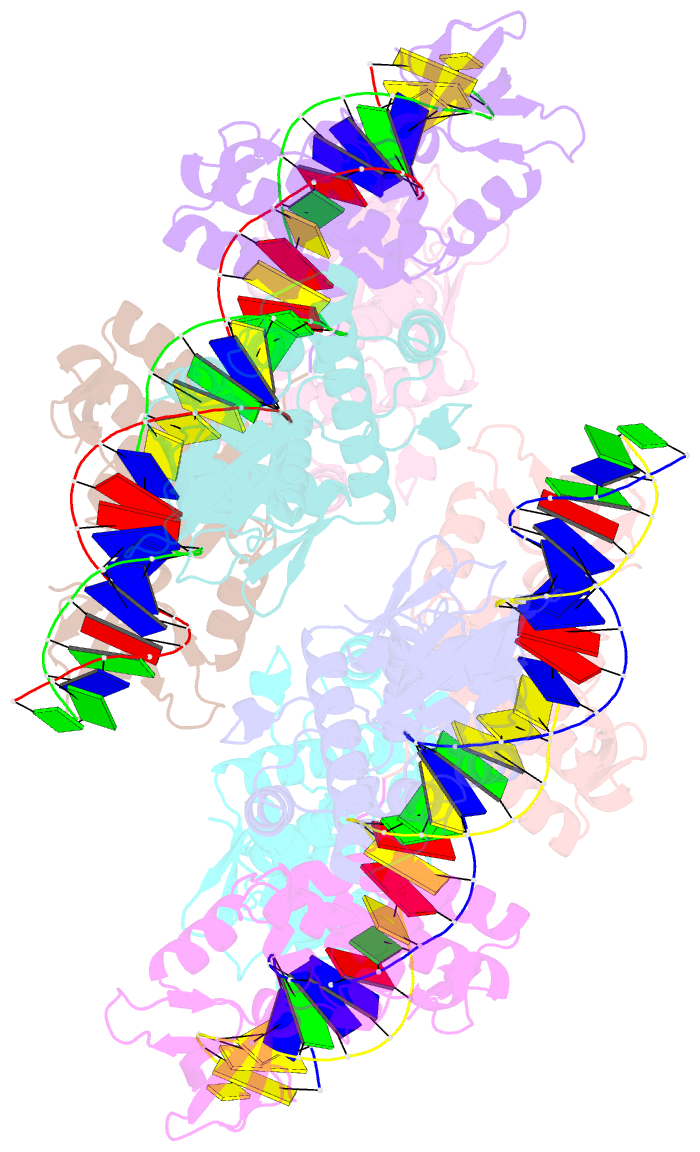
Summary information and primary citation
- PDB-id
- 1u8r; SNAP-derived features in text and JSON formats;
DNAproDB
- Class
- metal-binding protein,transcription-DNA
- Method
- X-ray (2.75 Å)
- Summary
- Crystal structure of an ider-DNA complex reveals a conformational change in activated ider for base-specific interactions
- Reference
- Wisedchaisri G, Holmes RK, Hol WGJ (2004): "Crystal Structure of an IdeR-DNA Complex Reveals a Conformational Change in Activated IdeR for Base-specific Interactions." J.Mol.Biol., 342, 1155-1169. doi: 10.1016/j.jmb.2004.07.083.
- Abstract
- The iron-dependent regulator (IdeR) is an essential protein in Mycobacterium tuberculosis that regulates iron uptake in this major pathogen. Under high iron concentrations, IdeR binds to several operator regions and represses transcription of target genes. Here, we report the first crystal structure of cobalt-activated IdeR bound to the mbtA-mbtB operator at 2.75 A resolution. IdeR binds to the DNA as a "double-dimer" complex with two dimers on opposite sides of the DNA duplex with the dimer axes deviating approximately 157 degrees. The asymmetric unit contains two such double-dimer complexes with a total molecular mass of 240 kDa. Two metal-binding sites are fully occupied with the SH3-like third domain adopting a "wedge" position to interact with the two other domains, and providing two ligands for the metal site 1 in all eight subunits per asymmetric unit. A putative sodium ion is observed to mediate interactions between Asp35 and DNA. There is a conformational change in the DNA-binding domain caused by a 6-9 degrees rotation of the helix-turn-helix motif with respect to the rest of the molecule upon binding to the DNA. Ser37 and Pro39 make specific interactions with conserved thymine bases while Gln43 makes non-specific contacts with different types of bases in different subunits. A "p1s2C3T4a5" base recognition pattern is proposed to be the basis for key interactions for each IdeR subunit with the DNA in the IdeR-DNA complex structure.


