Summary information and primary citation
- PDB-id
- 1xc8; SNAP-derived features in text and JSON formats;
DNAproDB
- Class
- hydrolase-DNA
- Method
- X-ray (1.95 Å)
- Summary
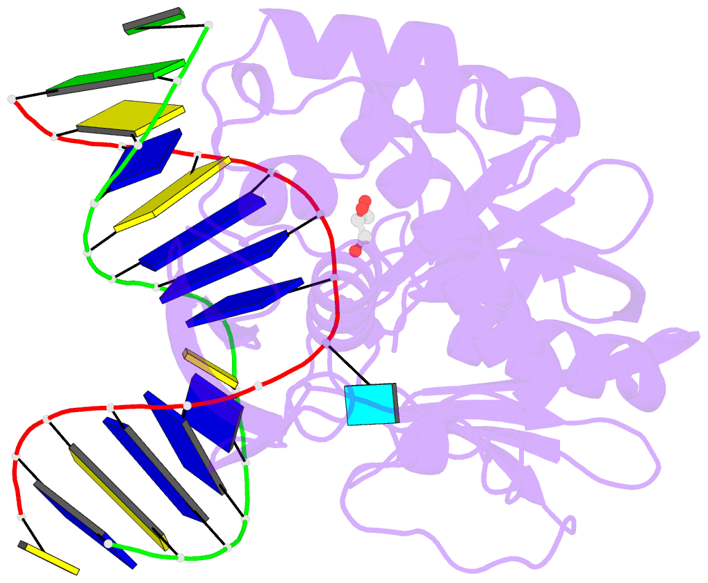
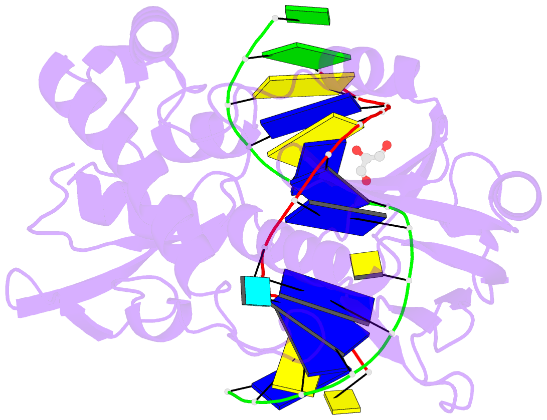
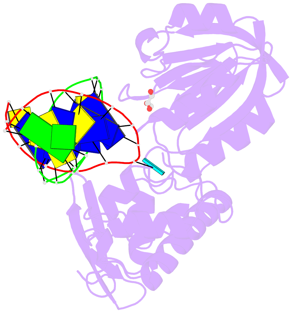
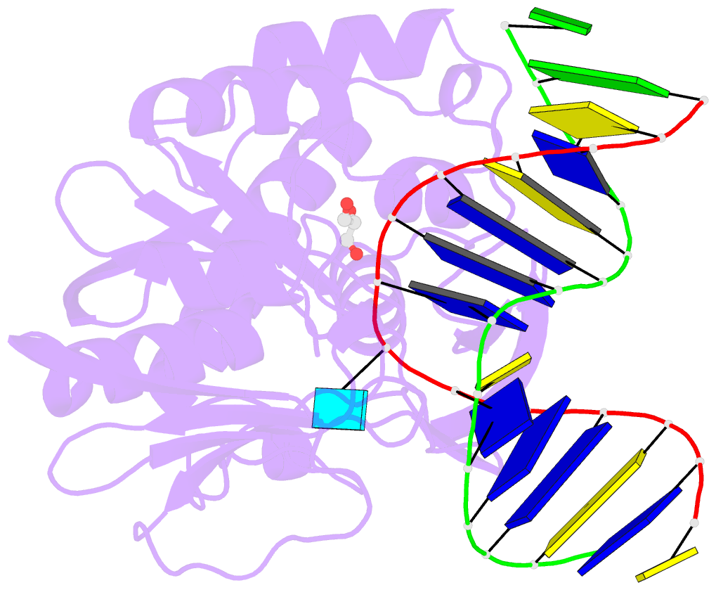
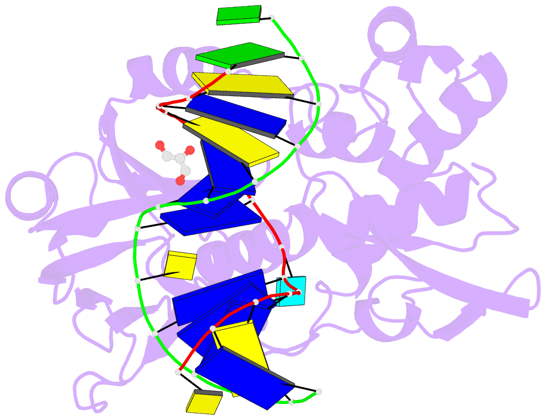
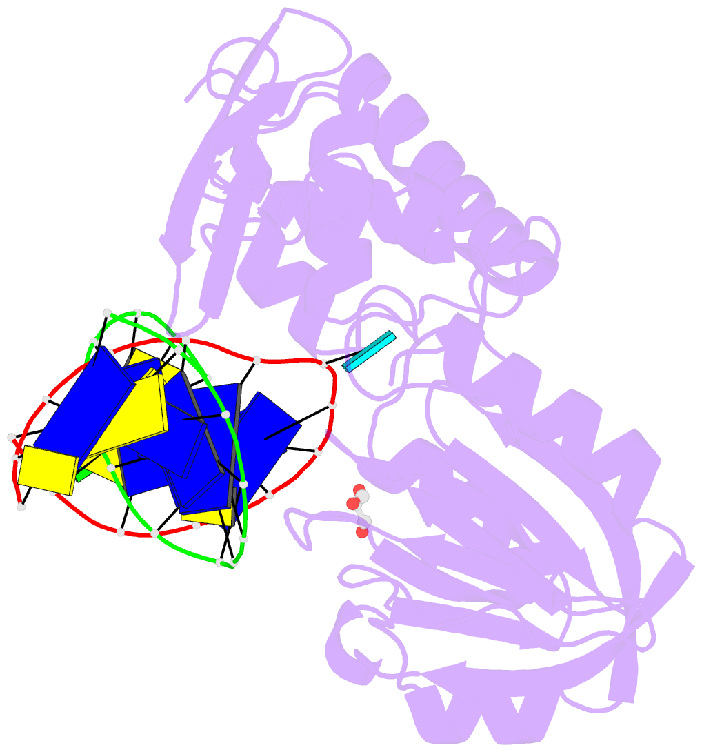
- Crystal structure complex between the wild-type lactococcus lactis fpg (mutm) and a fapy-dg containing DNA
- Reference
- Coste F, Ober M, Carell T, Boiteux S, Zelwer C, Castaing B (2004): "Structural basis for the recognition of the FapydG lesion (2,6-diamino-4-hydroxy-5-formamidopyrimidine) by formamidopyrimidine-DNA glycosylase." J.Biol.Chem., 279, 44074-44083. doi: 10.1074/jbc.M405928200.
- Abstract
- Formamidopyrimidine-DNA glycosylase (Fpg) is a DNA repair enzyme that excises oxidized purines such as 7,8-dihydro-8-oxoguanine (8-oxoG) and 2,6-diamino-4-hydroxy-5-formamidopyrimidine (FapyG) from damaged DNA. Here, we report the crystal structure of the Fpg protein from Lactococcus lactis (LlFpg) bound to a carbocyclic FapydG (cFapydG)-containing DNA. The structure reveals that Fpg stabilizes the cFapydG nucleoside into an extrahelical conformation inside its substrate binding pocket. In contrast to the recognition of the 8-oxodG lesion, which is bound with the glycosidic bond in a syn conformation, the cFapydG lesion displays in the complex an anti conformation. Furthermore, Fpg establishes interactions with all the functional groups of the FapyG base lesion, which can be classified in two categories: (i) those specifying a purine-derived lesion (here a guanine) involved in the Watson-Crick face recognition of the lesion and probably contributing to an optimal orientation of the pyrimidine ring moiety in the binding pocket and (ii) those specifying the imidazole ring-opened moiety of FapyG and probably participating also in the rotameric selection of the FapydG nucleobase. These interactions involve strictly conserved Fpg residues and structural water molecules mediated interactions. The significant differences between the Fpg recognition modes of 8-oxodG and FapydG provide new insights into the Fpg substrate specificity.