Summary information and primary citation
- PDB-id
- 1xi1; SNAP-derived features in text and JSON formats;
DNAproDB
- Class
- transferase-DNA
- Method
- X-ray (2.2 Å)
- Summary
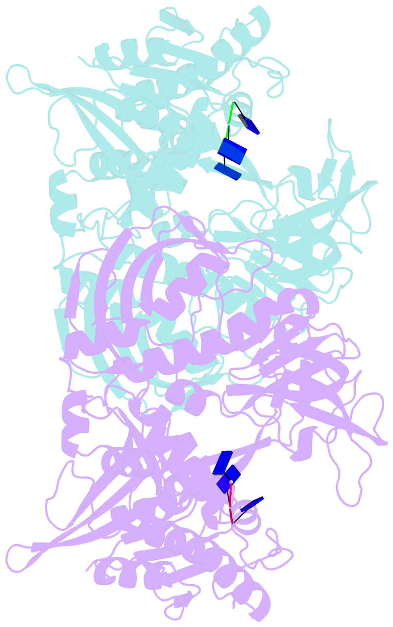
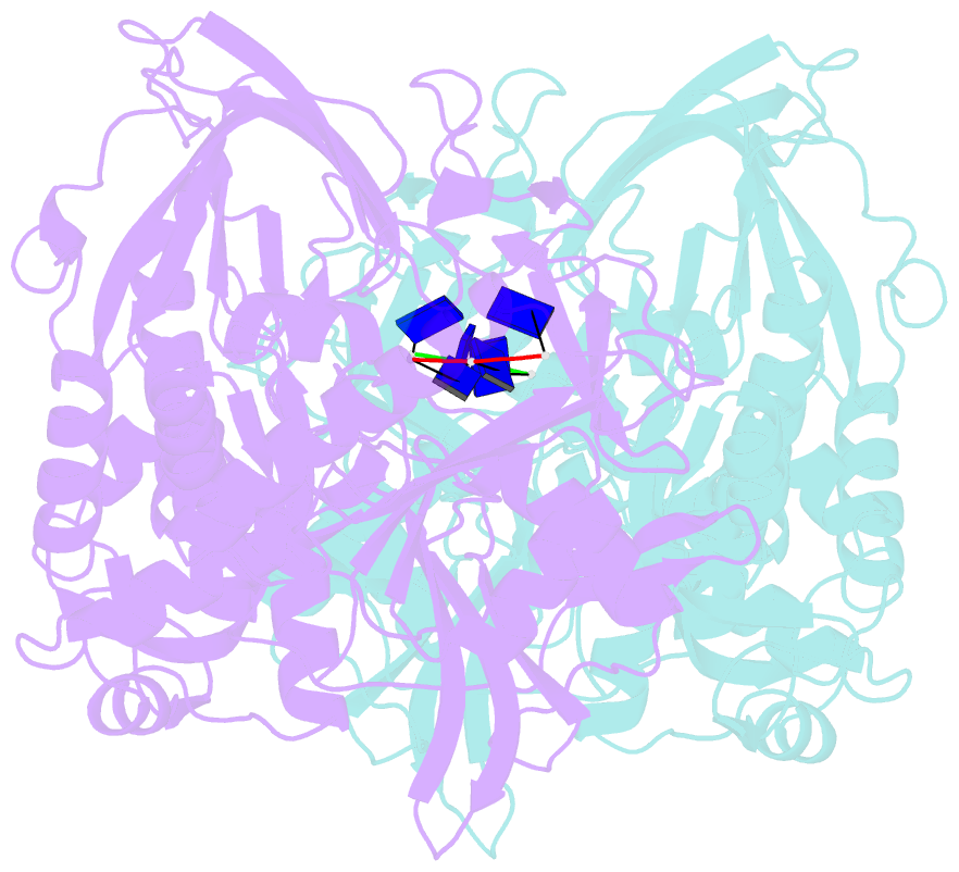
- Phi29 DNA polymerase ssDNA complex, monoclinic crystal form
- Reference
- Wang J, Kamtekar S, Berman AJ, Steitz TA (2005): "Correction of X-ray intensities from single crystals containing lattice-translocation defects." Acta Crystallogr.,Sect.D, 61, 67-74. doi: 10.1107/S0907444904026721.
- Abstract
- In 1954, Howells and colleagues described an unusual diffraction pattern from imidazole methemoglobin crystals caused by lattice-translocation defects. In these crystals, two identical lattices coexist as a single coherent mosaic block, but are translated by a fixed vector with respect to each other. The observed structure is a weighted sum of the two identical but translated structures, one from each lattice; the observed structure factors are a weighted vector sum of the two structure factors with identical unit amplitudes but shifted phases. A general procedure is described to obtain the unit amplitudes of observed structure factors from a realigned single lattice through an X-ray intensity correction. An application of this procedure is made to determine the crystal structure of phi29 DNA polymerase at 2.2 A resolution using multiple isomorphous replacement and multiwavelength anomalous dispersion methods.