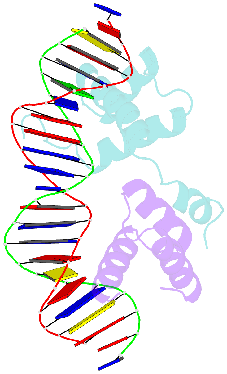
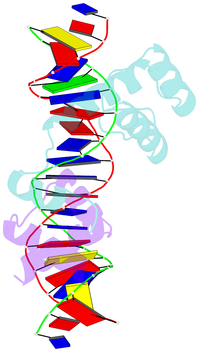
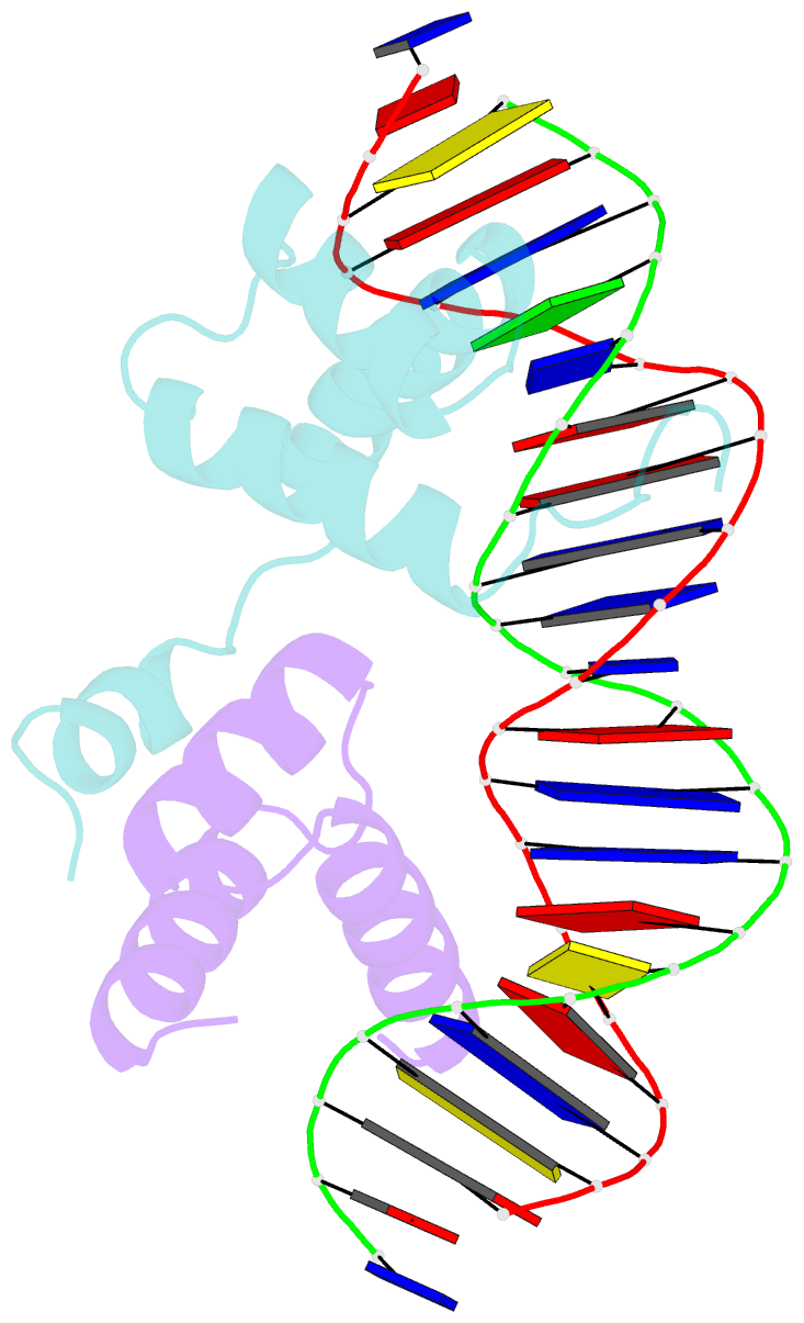
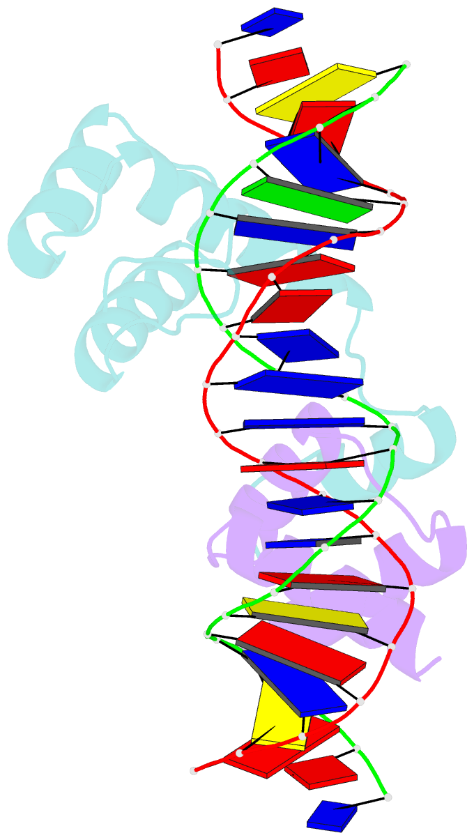
Summary information and primary citation
- PDB-id
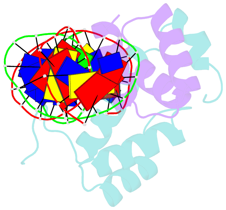
- 1yrn; SNAP-derived features in text and JSON formats;
DNAproDB
- Class
- DNA binding protein-DNA
- Method
- X-ray (2.5 Å)
- Summary
- Crystal structure of the mata1-matalpha2 homeodomain heterodimer bound to DNA
- Reference
- Li T, Stark MR, Johnson AD, Wolberger C (1995): "Crystal structure of the MATa1/MAT alpha 2 homeodomain heterodimer bound to DNA." Science, 270, 262-269.
- Abstract
- The Saccharomyces cerevisiae MATa1 and MAT alpha 2 homeodomain proteins, which play a role in determining yeast cell type, form a heterodimer that binds DNA and represses transcription in a cell type-specific manner. Whereas the alpha 2 and a1 proteins on their own have only modest affinity for DNA, the a1/alpha 2 heterodimer binds DNA with high specificity and affinity. The three-dimensional crystal structure of the a1/alpha 2 homeodomain heterodimer bound to DNA was determined at a resolution of 2.5 A. The a1 and alpha 2 homeodomains bind in a head-to-tail orientation, with heterodimer contacts mediated by a 16-residue tail located carboxyl-terminal to the alpha 2 homeodomain. This tail becomes ordered in the presence of a1, part of it forming a short amphipathic helix that packs against the a1 homeodomain between helices 1 and 2. A pronounced 60 degree bend is induced in the DNA, which makes possible protein-protein and protein-DNA contacts that could not take place in a straight DNA fragment. Complex formation mediated by flexible protein-recognition peptides attached to stably folded DNA binding domains may prove to be a general feature of the architecture of other classes of eukaryotic transcriptional regulators.