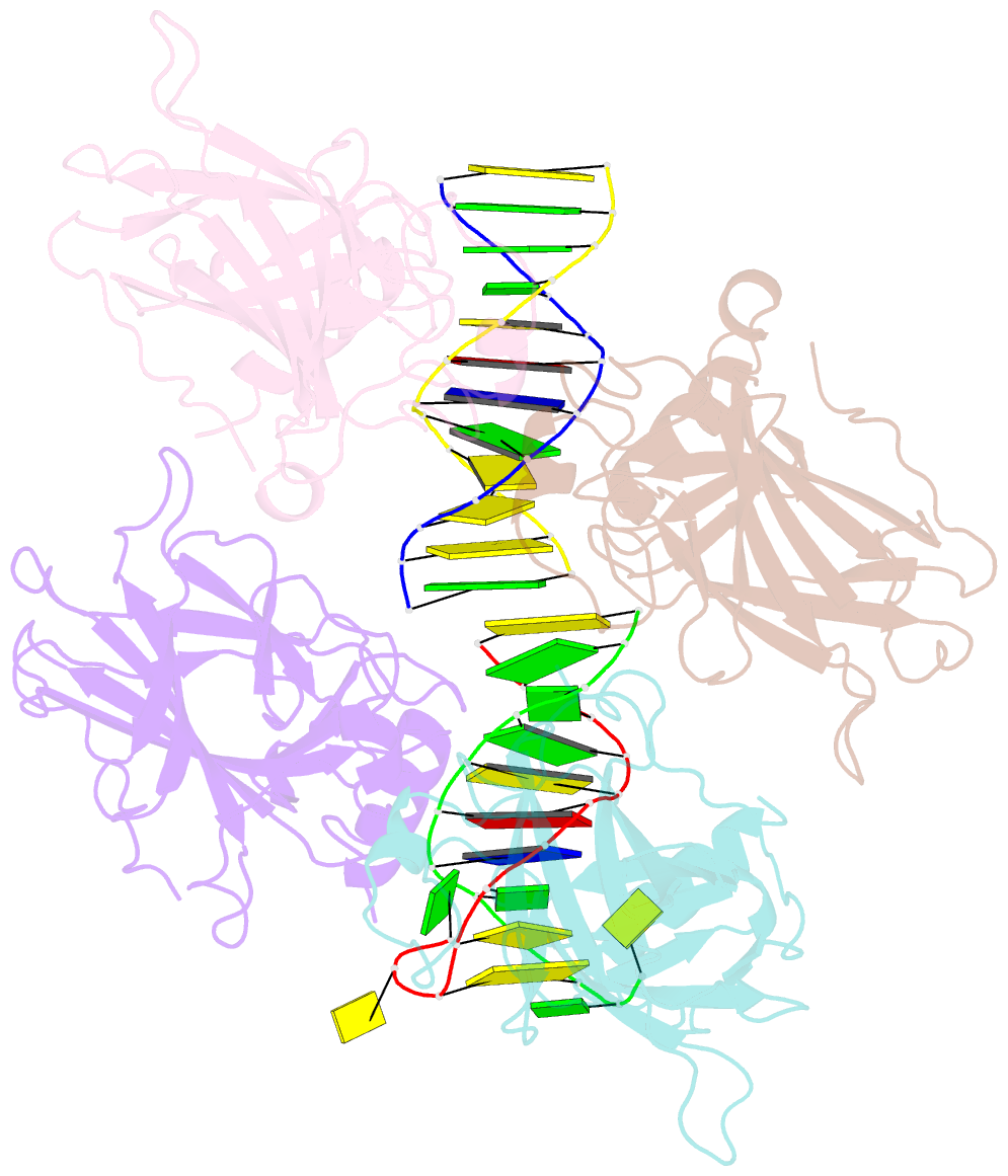
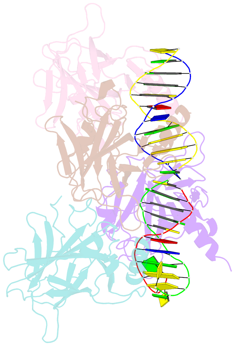
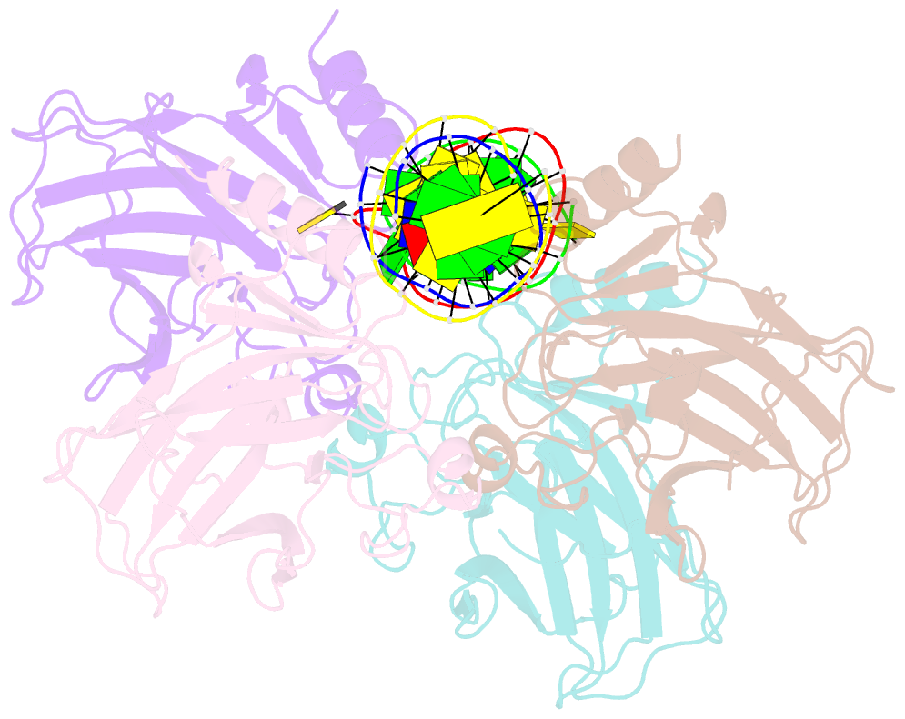
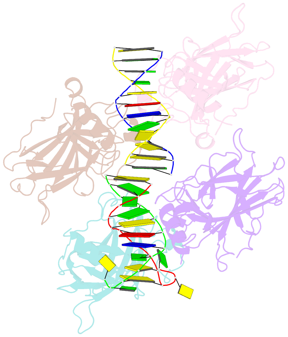
Summary information and primary citation
- PDB-id
- 2ac0; SNAP-derived features in text and JSON formats;
DNAproDB
- Class
- apoptosis-DNA
- Method
- X-ray (1.8 Å)
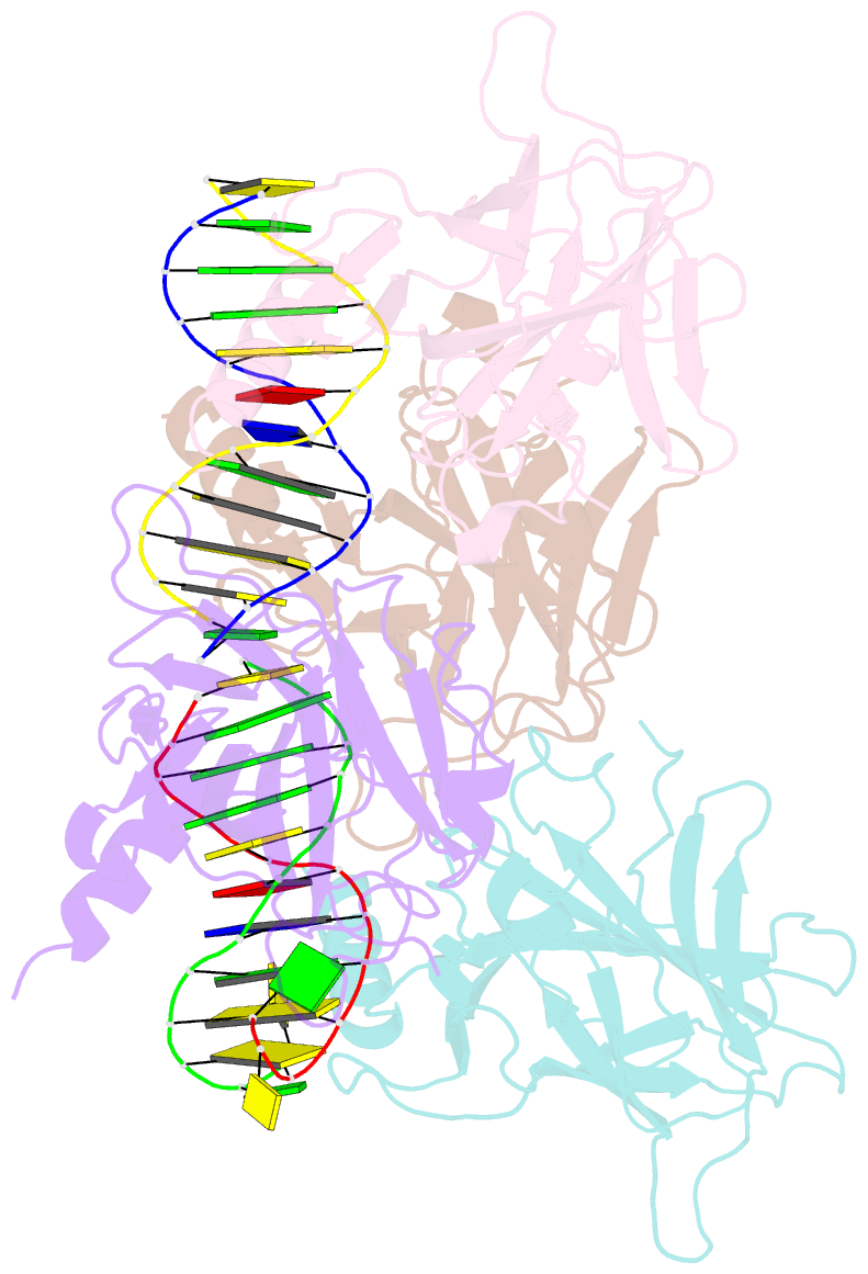
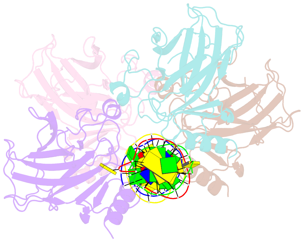
- Summary
- Structural basis of DNA recognition by p53 tetramers (complex i)
- Reference
- Kitayner M, Rozenberg H, Kessler N, Rabinovich D, Shaulov L, Haran TE, Shakked Z (2006): "Structural Basis of DNA Recognition by p53 Tetramers." Mol.Cell, 22, 741-753. doi: 10.1016/j.molcel.2006.05.015.
- Abstract
- The tumor-suppressor protein p53 is among the most effective of the cell's natural defenses against cancer. In response to cellular stress, p53 binds as a tetramer to diverse DNA targets containing two decameric half-sites, thereby activating the expression of genes involved in cell-cycle arrest or apoptosis. Here we present high-resolution crystal structures of sequence-specific complexes between the core domain of human p53 and different DNA half-sites. In all structures, four p53 molecules self-assemble on two DNA half-sites to form a tetramer that is a dimer of dimers, stabilized by protein-protein and base-stacking interactions. The protein-DNA interface varies as a function of the specific base sequence in correlation with the measured binding affinities of the complexes. The new data establish a structural framework for understanding the mechanisms of specificity, affinity, and cooperativity of DNA binding by p53 and suggest a model for its regulation by regions outside the sequence-specific DNA binding domain.