Summary information and primary citation
- PDB-id
- 2axy; SNAP-derived features in text and JSON formats;
DNAproDB
- Class
- DNA binding protein-DNA
- Method
- X-ray (1.7 Å)
- Summary
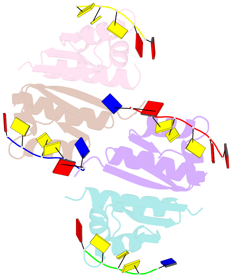
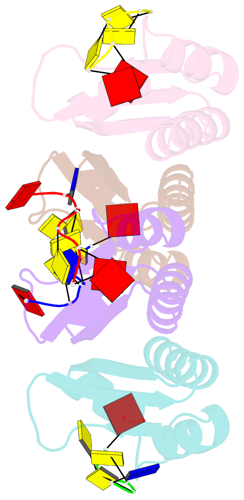
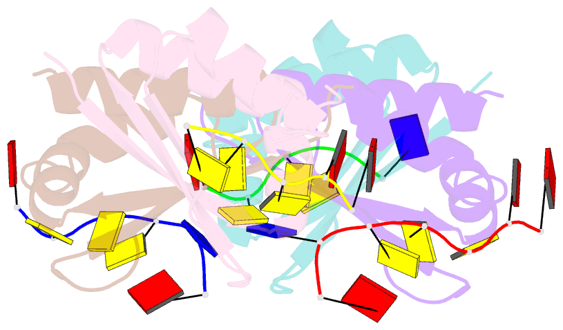
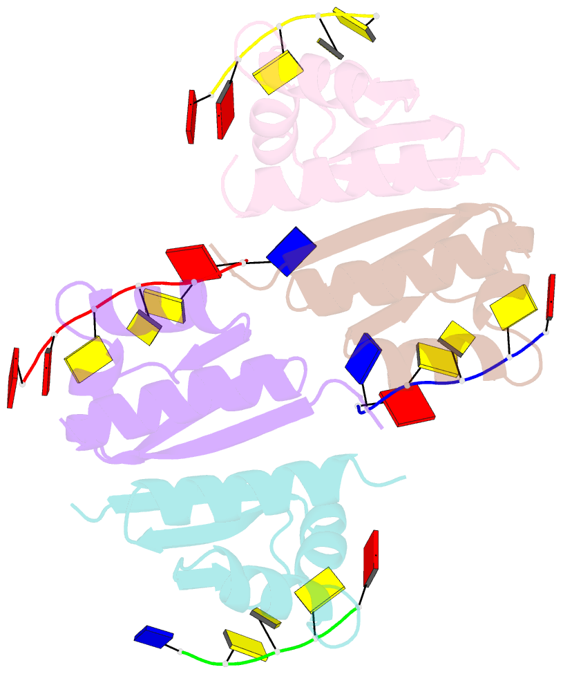
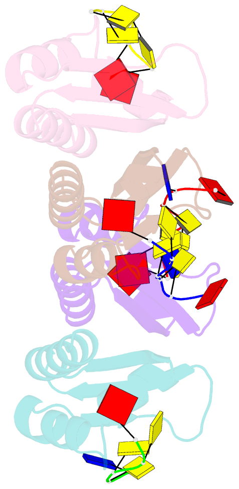
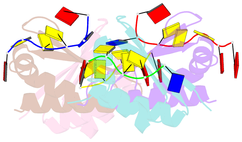
- Crystal structure of kh1 domain of human poly(c)-binding protein-2 with c-rich strand of human telomeric DNA
- Reference
- Du Z, Lee JK, Tjhen R, Li S, Pan H, Stroud RM, James TL (2005): "Crystal Structure of the First KH Domain of Human Poly(C)-binding Protein-2 in Complex with a C-rich Strand of Human Telomeric DNA at 1.7 A." J.Biol.Chem., 280, 38823-38830. doi: 10.1074/jbc.M508183200.
- Abstract
- Recognition of poly(C) DNA and RNA sequences in mammalian cells is achieved by a subfamily of the KH (hnRNP K homology) domain-containing proteins known as poly(C)-binding proteins (PCBPs). To reveal the molecular basis of poly(C) sequence recognition, we have determined the crystal structure, at 1.7-A resolution, of PCBP2 KH1 in complex with a 7-nucleotide DNA sequence (5'-AACCCTA-3') corresponding to one repeat of the human C-rich strand telomeric DNA. The protein-DNA interaction is mediated by the combination of several stabilizing forces including hydrogen bonding, electrostatic interactions, van der Waals contacts, and shape complementarities. Specific recognition of the three cytosine residues is realized by a dense network of hydrogen bonds involving the side chains of two conserved lysines and one glutamic acid. The co-crystal structure also reveals a protein-protein dimerization interface of PCBP2 KH1 located on the opposite side of the protein from the DNA binding groove. Numerous stabilizing protein-protein interactions, including hydrophobic contacts, stacking of aromatic side chains, and a large number of hydrogen bonds, indicate that the protein-protein interaction interface is most likely genuine. Interaction of PCBP2 KH1 with the C-rich strand of human telomeric DNA suggests that PCBPs may participate in mechanisms involved in the regulation of telomere/telomerase functions.