Summary information and primary citation
- PDB-id
- 2b9s; SNAP-derived features in text and JSON formats;
DNAproDB
- Class
- isomerase-DNA
- Method
- X-ray (2.27 Å)
- Summary
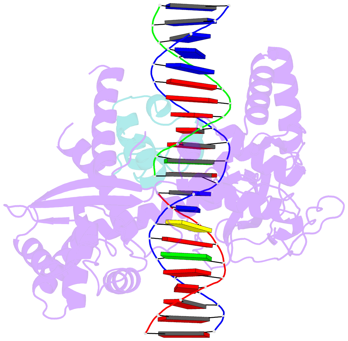
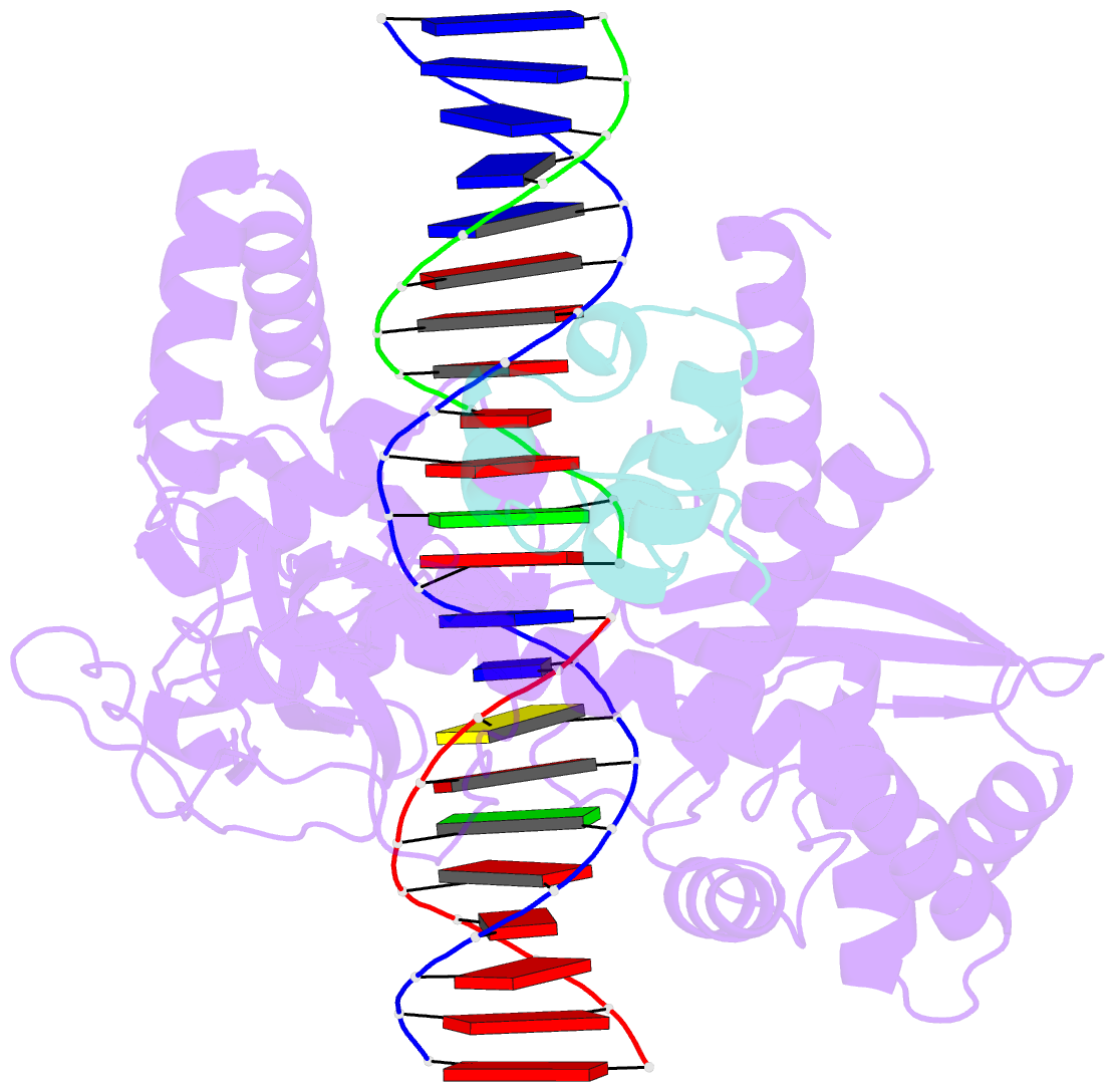
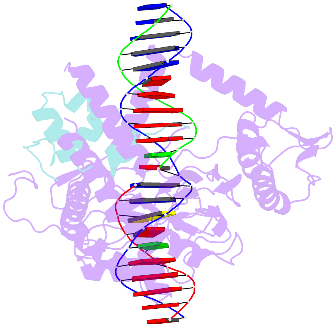
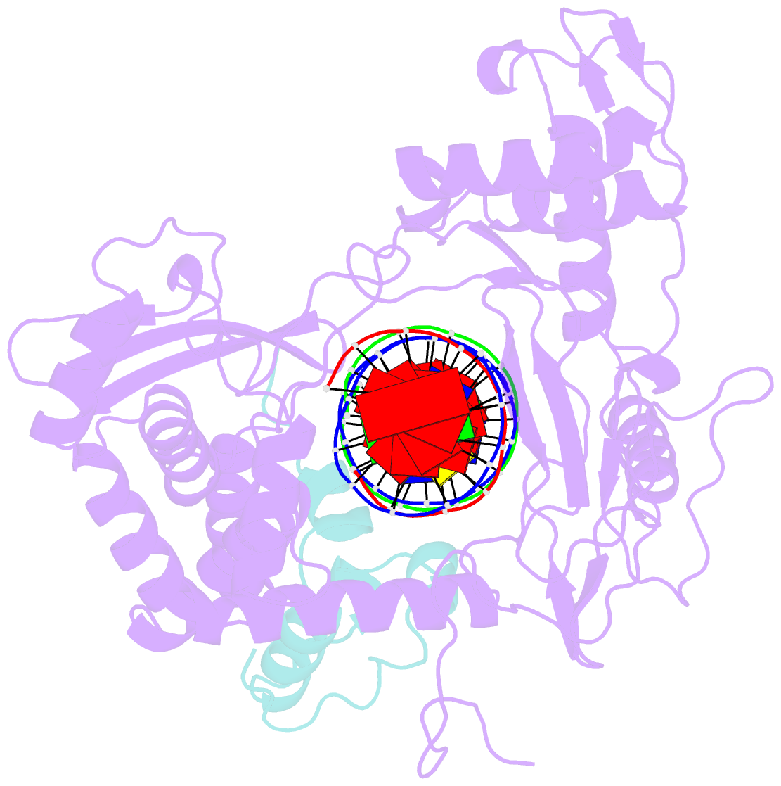
- Crystal structure of heterodimeric l. donovani topoisomerase i-vanadate-DNA complex
- Reference
- Davies DR, Mushtaq A, Interthal H, Champoux JJ, Hol WG (2006): "The Structure of the Transition State of the Heterodimeric Topoisomerase I of Leishmania donovani as a Vanadate Complex with Nicked DNA." J.Mol.Biol., 357, 1202-1210. doi: 10.1016/j.jmb.2006.01.022.
- Abstract
- Type IB topoisomerases are essential enzymes that are responsible for relaxing superhelical tension in DNA by forming a transient covalent nick in one strand of the DNA duplex. Topoisomerase I is a target for anti-cancer drugs such as camptothecin, and these drugs also target the topoisomerases I in pathogenic trypanosomes including Leishmania species and Trypanosoma brucei. Most eukaryotic enzymes, including human topoisomerase I, are monomeric. However, for Leishmania donovani, the DNA-binding activity and the majority of residues involved in catalysis are located in a large subunit, designated TOP1L, whereas the catalytic tyrosine residue responsible for covalent attachment to DNA is located in a smaller subunit, called TOP1S. Here, we present the 2.27A crystal structure of an active truncated L.donovani TOP1L/TOP1S heterodimer bound to nicked double-stranded DNA captured as a vanadate complex. The vanadate forms covalent linkages between the catalytic tyrosine residue of the small subunit and the nicked ends of the scissile DNA strand, mimicking the previously unseen transition state of the topoisomerase I catalytic cycle. This structure fills a critical gap in the existing ensemble of topoisomerase I structures and provides crucial insights into the catalytic mechanism.