Summary information and primary citation
- PDB-id
- 2bbv; SNAP-derived features in text and JSON formats;
DNAproDB
- Class
- virus-RNA
- Method
- X-ray (2.8 Å)
- Summary
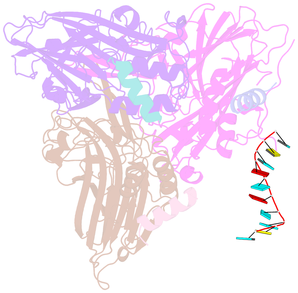
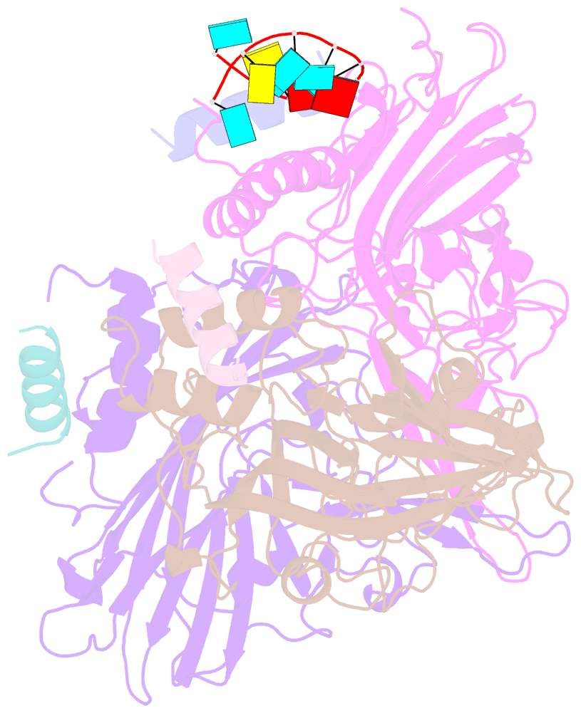
- The refined three-dimensional structure of an insect virus at 2.8 angstroms resolution
- Reference
- Wery JP, Reddy VS, Hosur MV, Johnson JE (1994): "The refined three-dimensional structure of an insect virus at 2.8 A resolution." J.Mol.Biol., 235, 565-586. doi: 10.1006/jmbi.1994.1014.
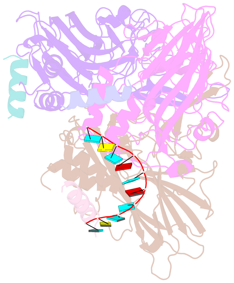
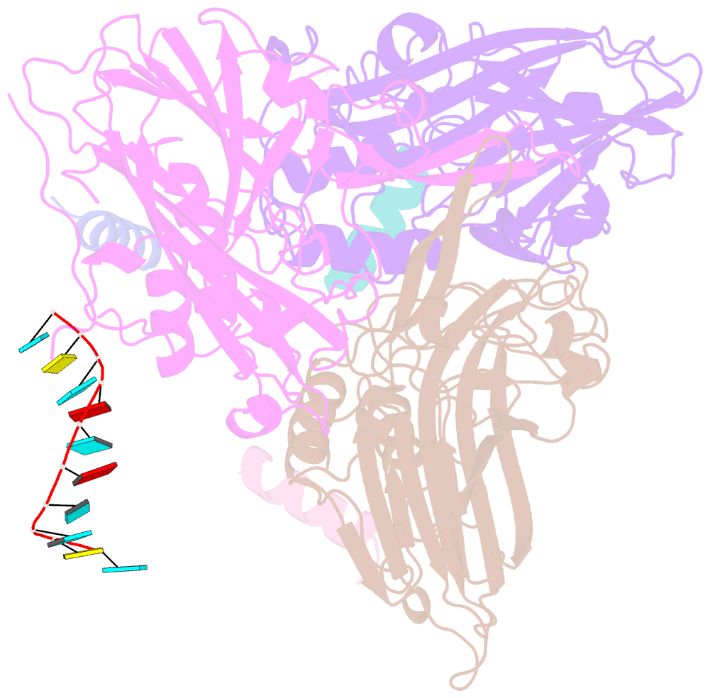
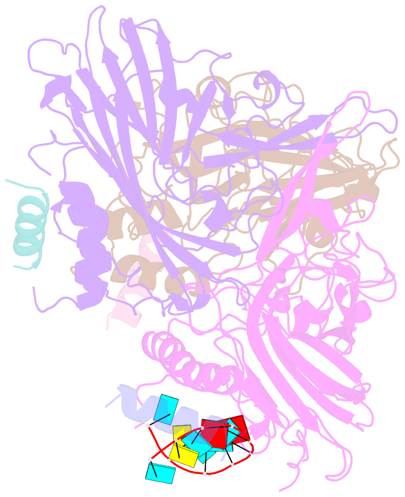
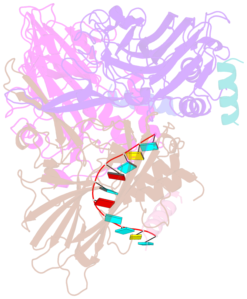
- Abstract
- The structure of the black beetle nodavirus has been refined at 2.8 A resolution by alternate use of restrained least-squares atomic coordinate refinement and phase refinement by real space averaging with the 5-fold non-crystallographic symmetry in the crystal. The coordinates were also refined by simulated annealing. The final R-factor for all data with I/sigma(I) > 4 was 22.1%. A total of 7692 atoms were refined in one icosahedral asymmetric unit which included 273 oxygen atoms of ordered water molecules. Three identical gene products of 407 amino acids form one icosahedral asymmetric unit. Each is located in a structurally unique position, identified as A, B or C, consistent with a T = 3 quasi equivalent lattice. Icosahedral pentamers are formed by A subunits while B and C subunits alternate about icosahedral 3-fold axes to form quasi hexamers. Five calcium ions are located within the icosahedral asymmetric unit and stabilize the quasi 3-fold related intersubunit contacts between A, B and C. The final model consists of coordinates for residues 56 to 379 of all three subunits and residues 20 to 31 from the C subunit only. Atom positions for the sugar-phosphate backbone were modeled for ten nucleotides close to the icosahedral 2-fold axes. Symmetry equivalent polyribonucleotides form a helical duplex at each icosahedral 2-fold axis. The three subunits display an eight-stranded beta-barrel fold, very similar to the subunit structures observed in most other icosahedral RNA viruses analyzed. Quasi equivalence is regulated by the ordered RNA and residues 20 to 31 in the C subunit to form a "flat inter subunit contact" at icosahedral 2-fold joints. The RNA and polypeptide are disordered at the quasi 2-fold joints and this results in a "bent inter subunit contact". Although similar quaternary structures were seen in T = 3 plant viruses studied, RNA did not play a role as a molecular switch in those structures. The autocatalytic, post assembly, cleavage of the initial gene product at residue Asn363/Ala364 to form a stable and infectious particle is probably the result of an acid catalyzed main-chain hydrolysis in which Asp75 is the proton donor. The reaction is initiated by assembly which places Asp75 in a hydrophobic environment created by quaternary interactions which raises its pK to 5.6. The region in which the reaction occurs is formed by an internal helical bundle that has not been seen in other virus structures.