Summary information and primary citation
- PDB-id
- 2bop; SNAP-derived features in text and JSON formats;
DNAproDB
- Class
- transcription-DNA
- Method
- X-ray (1.7 Å)
- Summary
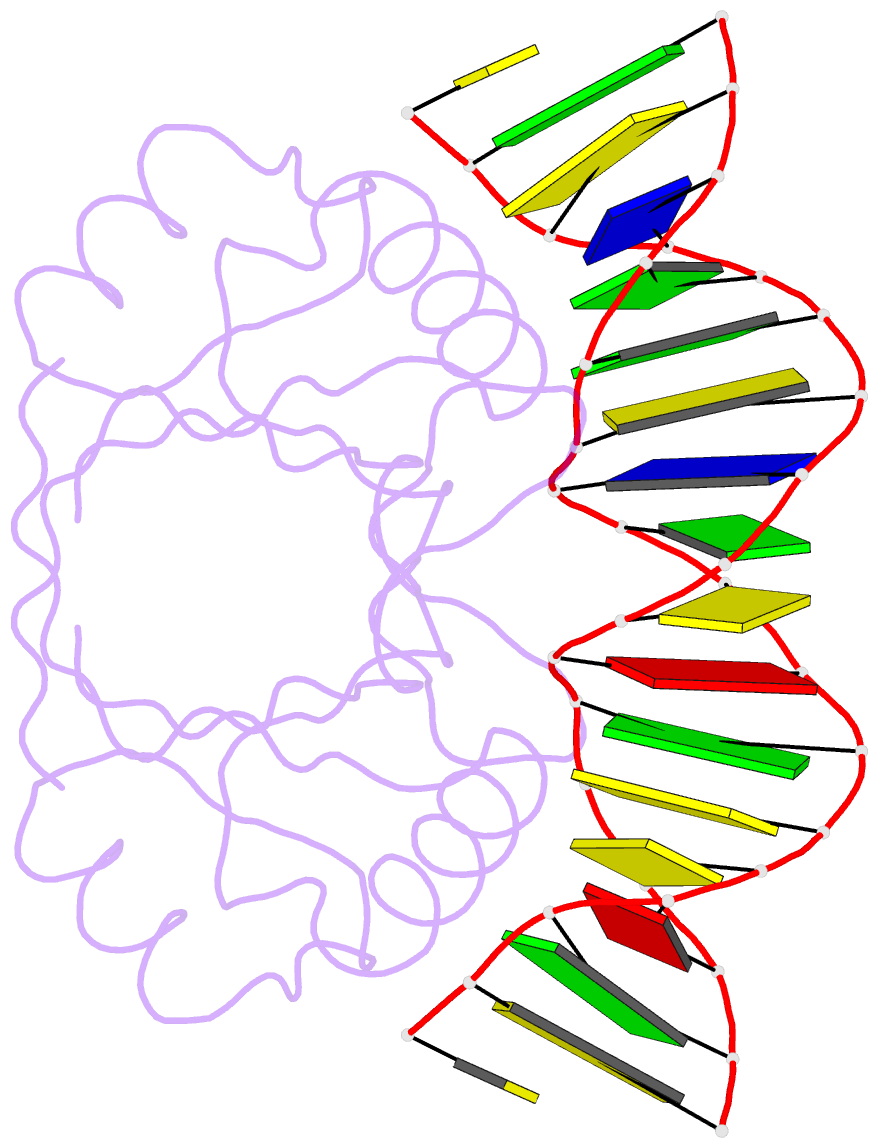
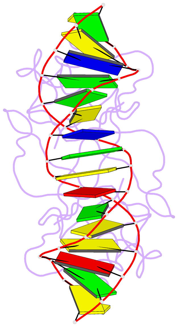
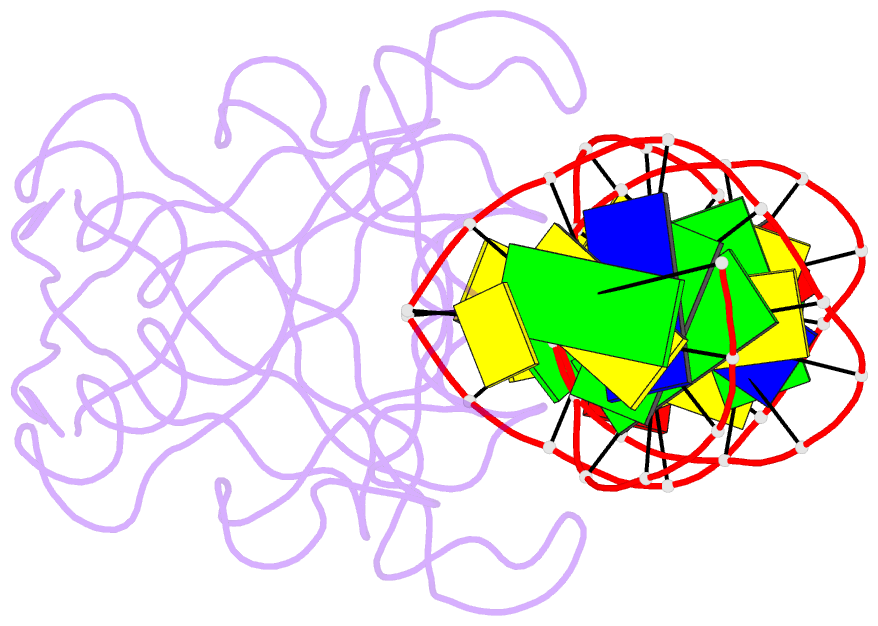
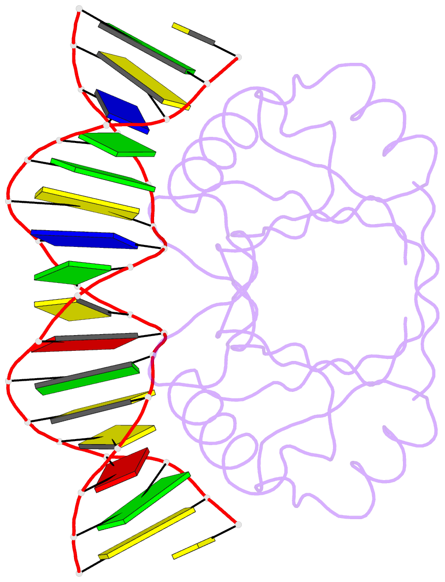
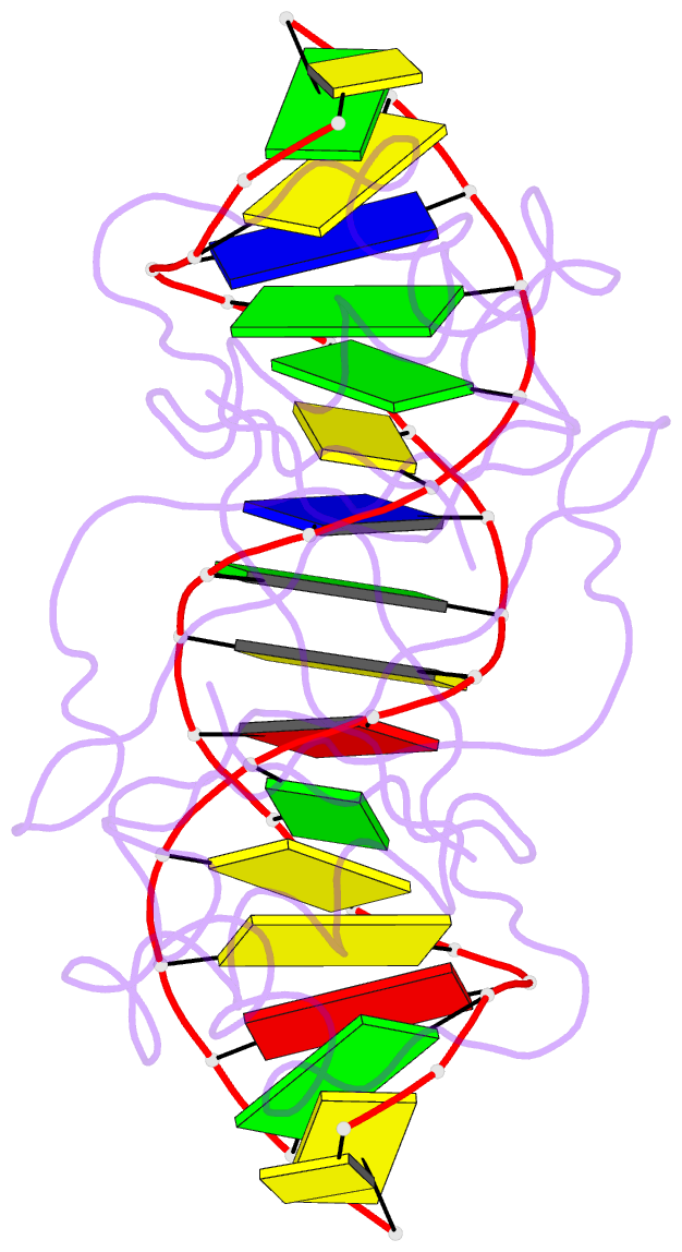
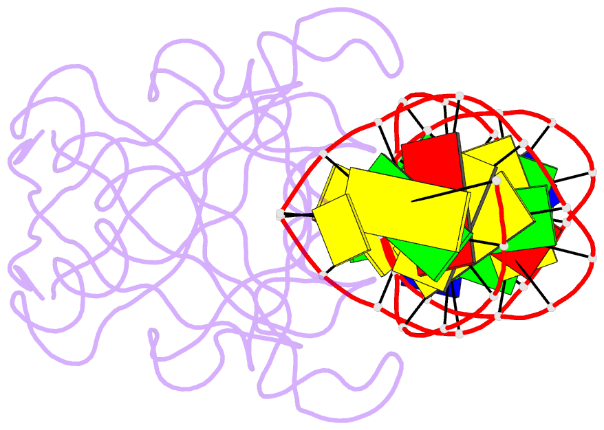
- Crystal structure at 1.7 angstroms of the bovine papillomavirus-1 e2 DNA-binding domain bound to its DNA target
- Reference
- Hegde RS, Grossman SR, Laimins LA, Sigler PB (1992): "Crystal structure at 1.7 A of the bovine papillomavirus-1 E2 DNA-binding domain bound to its DNA target." Nature, 359, 505-512. doi: 10.1038/359505a0.
- Abstract
- The dominant transcriptional regulator of the papillomaviruses, E2, binds to its specific DNA target through a previously unobserved dimeric antiparallel beta-barrel. The DNA is severely but smoothly bent over the barrel by the interaction of successive major grooves with a pair of symmetrically disposed alpha-helices. The specific interface is an 'interwoven' network of interactions where the identifying base pairs of the target contact more than one amino-acid side chain and the discriminating amino acids interact with more than one base pair.