Summary information and primary citation
- PDB-id
- 2c7r; SNAP-derived features in text and JSON formats;
DNAproDB
- Class
- transferase-DNA
- Method
- X-ray (1.9 Å)
- Summary
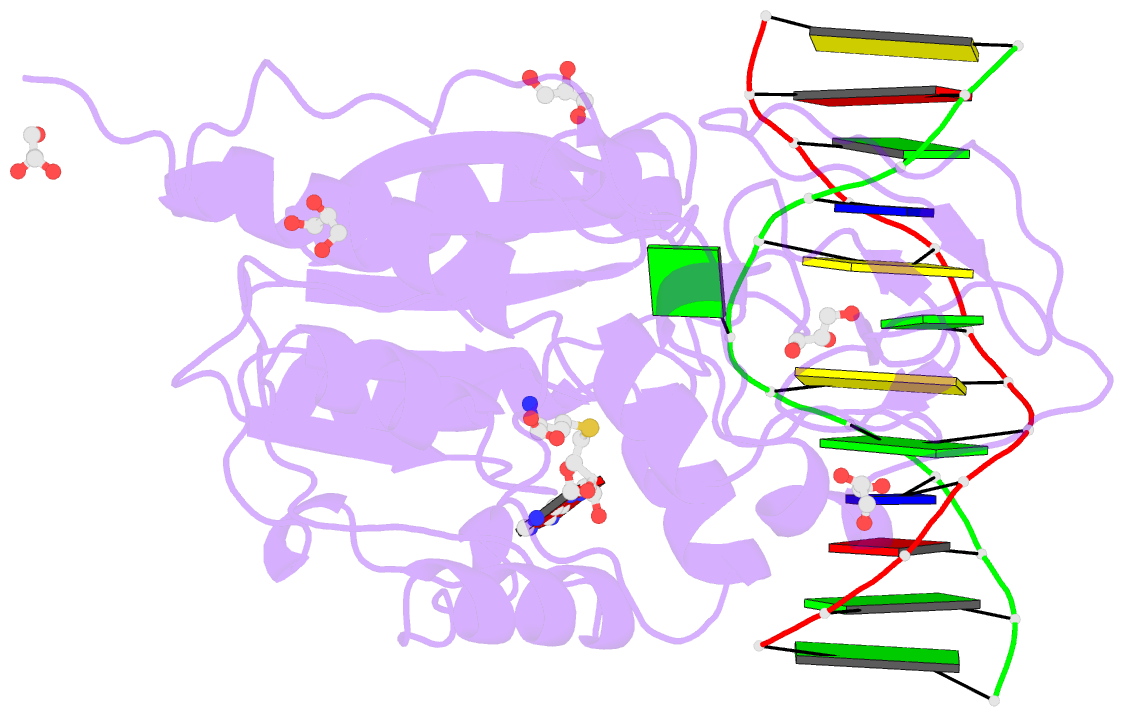
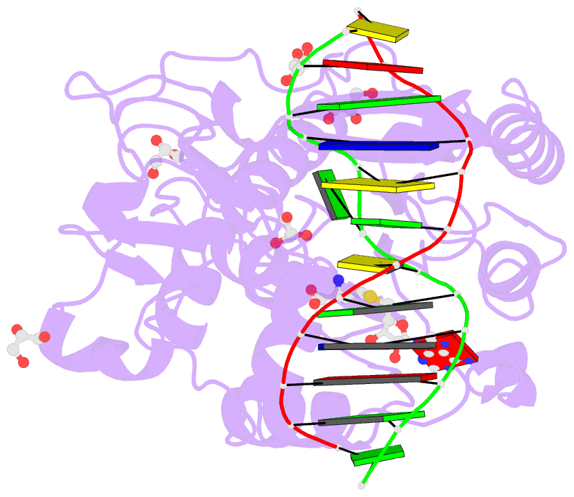
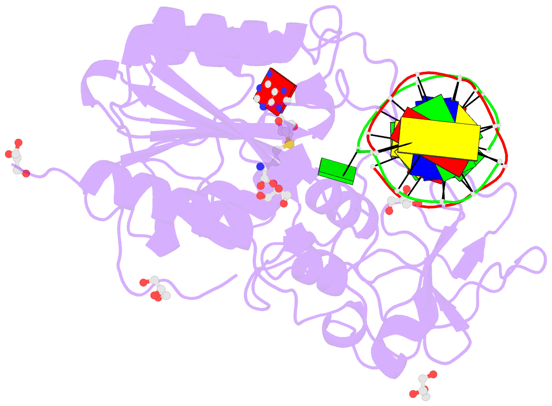
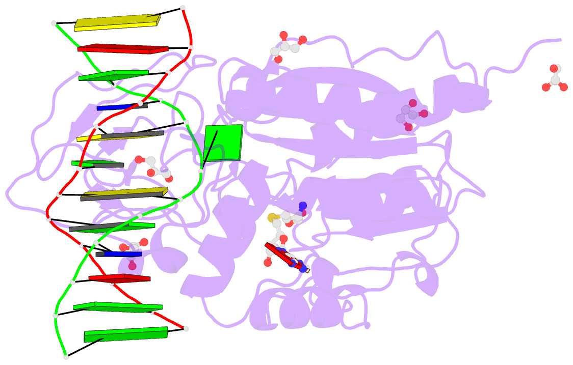
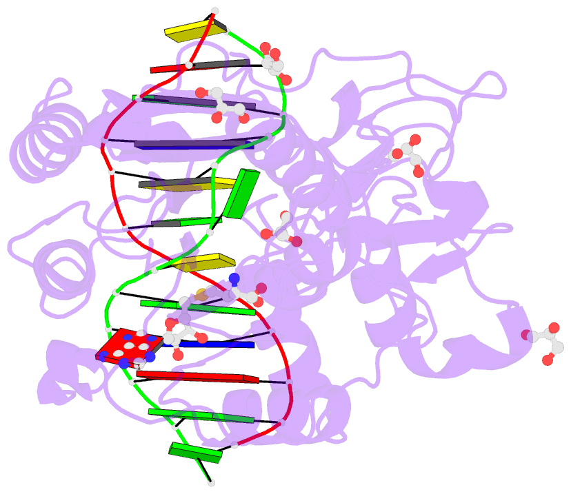
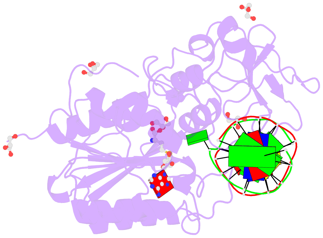
- Hhai DNA methyltransferase (t250g mutant) complex with oligonucleotide containing 2-aminopurine as a target base (gpgc:gmgc) and sah
- Reference
- Neely RK, Daujotyte D, Grazulis S, Magennis SW, Dryden DTF, Klimasauskas S, Jones AC (2005): "Time-Resolved Fluorescence of 2-Aminopurine as a Probe of Base Flipping in M.HhaI-DNA Complexes." Nucleic Acids Res., 33, 6953. doi: 10.1093/NAR/GKI995.
- Abstract
- DNA base flipping is an important mechanism in molecular enzymology, but its study is limited by the lack of an accessible and reliable diagnostic technique. A series of crystalline complexes of a DNA methyltransferase, M.HhaI, and its cognate DNA, in which a fluorescent nucleobase analogue, 2-aminopurine (AP), occupies defined positions with respect the target flipped base, have been prepared and their structures determined at higher than 2 A resolution. From time-resolved fluorescence measurements of these single crystals, we have established that the fluorescence decay function of AP shows a pronounced, characteristic response to base flipping: the loss of the very short (approximately 100 ps) decay component and the large increase in the amplitude of the long (approximately 10 ns) component. When AP is positioned at sites other than the target site, this response is not seen. Most significantly, we have shown that the same clear response is apparent when M.HhaI complexes with DNA in solution, giving an unambiguous signal of base flipping. Analysis of the AP fluorescence decay function reveals conformational heterogeneity in the DNA-enzyme complexes that cannot be discerned from the present X-ray structures.