Summary information and primary citation
- PDB-id
- 2dem; SNAP-derived features in text and JSON formats;
DNAproDB
- Class
- hydrolase-DNA
- Method
- X-ray (1.95 Å)
- Summary
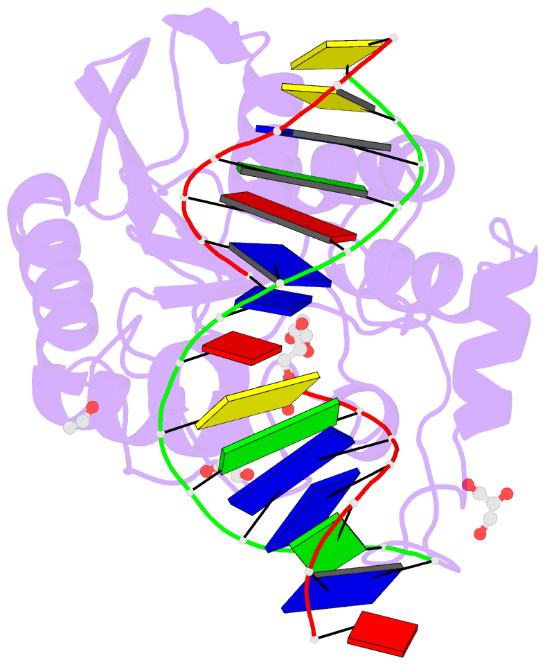
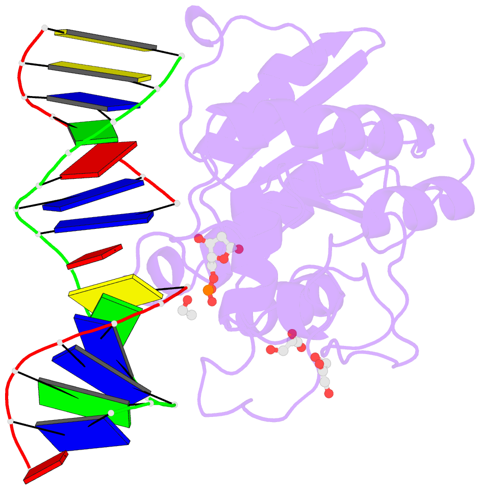
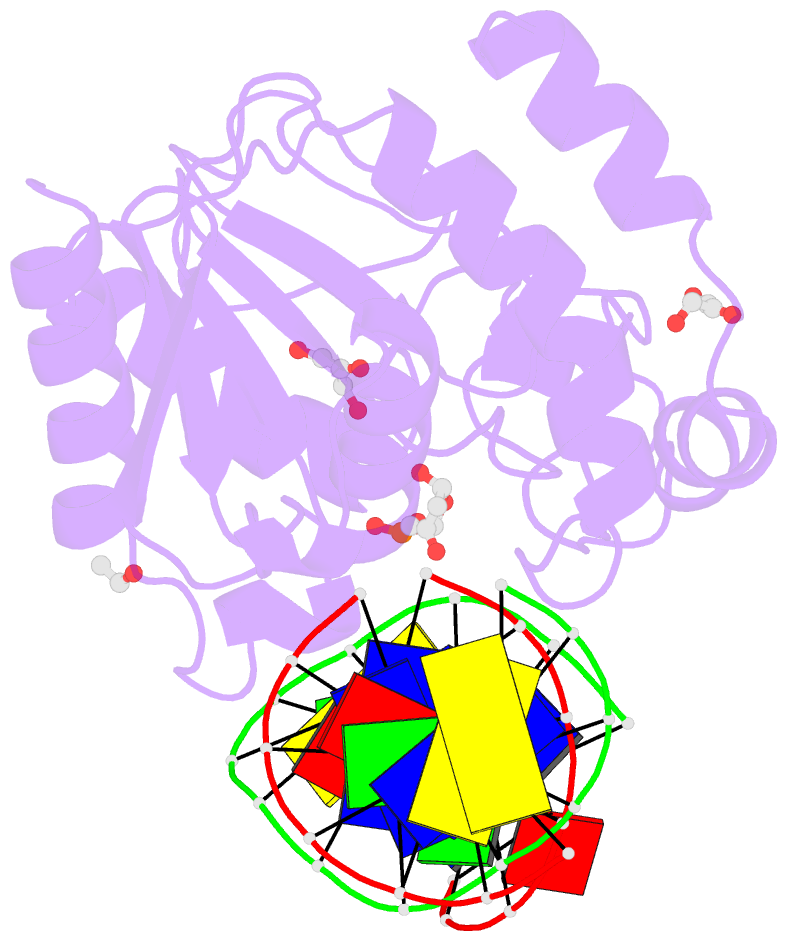
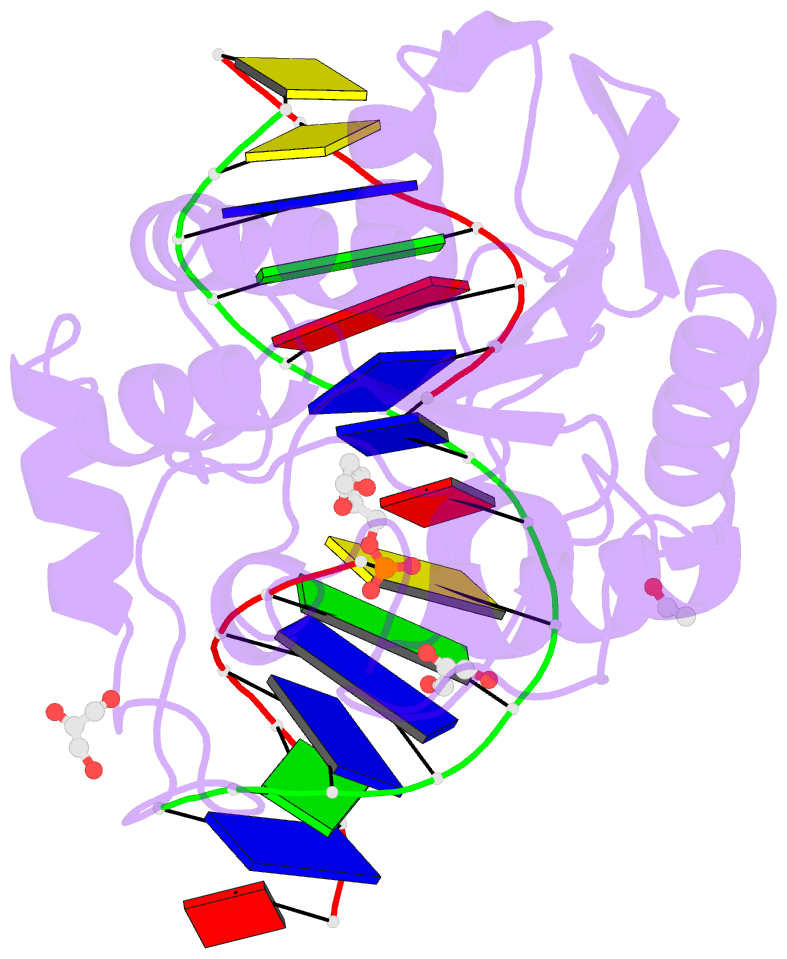
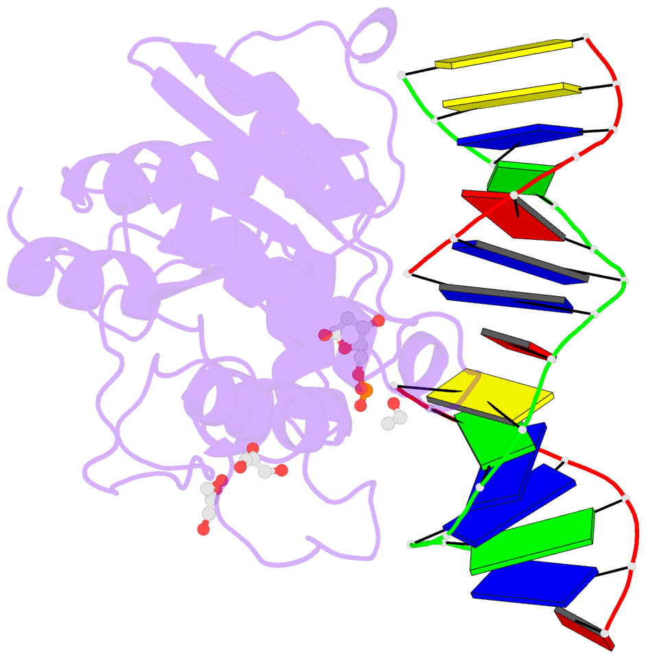
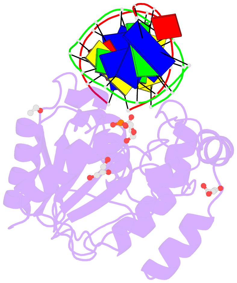
- Crystal structure of uracil-DNA glycosylase in complex with ap:a containing DNA
- Reference
- Kosaka H, Hoseki J, Nakagawa N, Kuramitsu S, Masui R (2007): "Crystal structure of family 5 uracil-DNA glycosylase bound to DNA." J.Mol.Biol., 373, 839-850. doi: 10.1016/j.jmb.2007.08.022.
- Abstract
- Uracil-DNA glycosylase (UDG) removes uracil generated by the deamination of cytosine or misincorporation of deoxyuridine monophosphate. Within the UDG superfamily, a fifth UDG family lacks a polar residue in the active-site motif, which mediates the hydrolysis of the glycosidic bond by activation of a water molecule in UDG families 1-4. We have determined the crystal structure of a novel family 5 UDG from Thermus thermophilus HB8 complexed with DNA containing an abasic site. The active-site structure suggests this enzyme uses both steric force and water activation for its excision reaction. A conserved asparagine residue acts as a ligand to the catalytic water molecule. The structure also implies that another water molecule acts as a barrier during substrate recognition. Based on no significant open-closed conformational change upon binding to DNA, we propose a "slide-in" mechanism for initial damage recognition.