Summary information and primary citation
- PDB-id
- 2fkc; SNAP-derived features in text and JSON formats;
DNAproDB
- Class
- hydrolase-DNA
- Method
- X-ray (2.39 Å)
- Summary
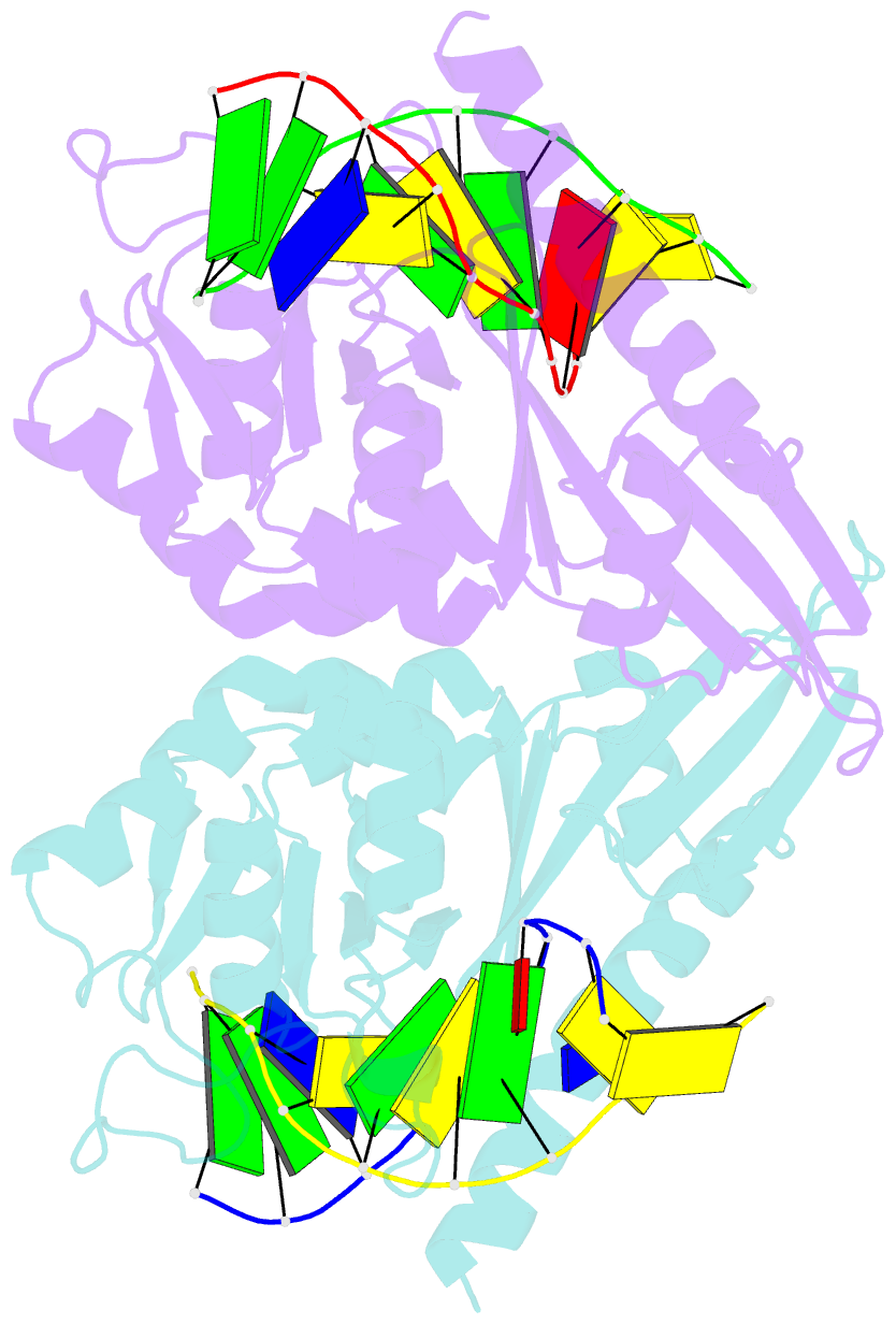
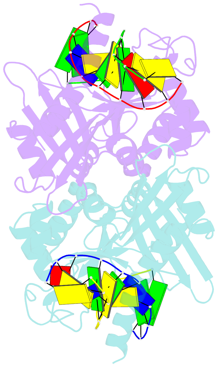
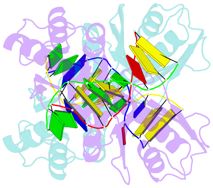
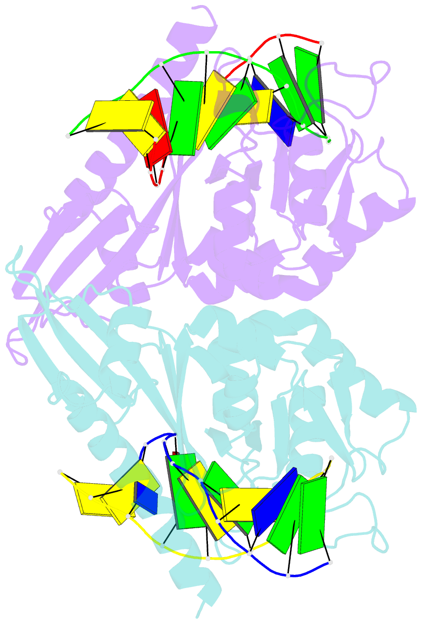
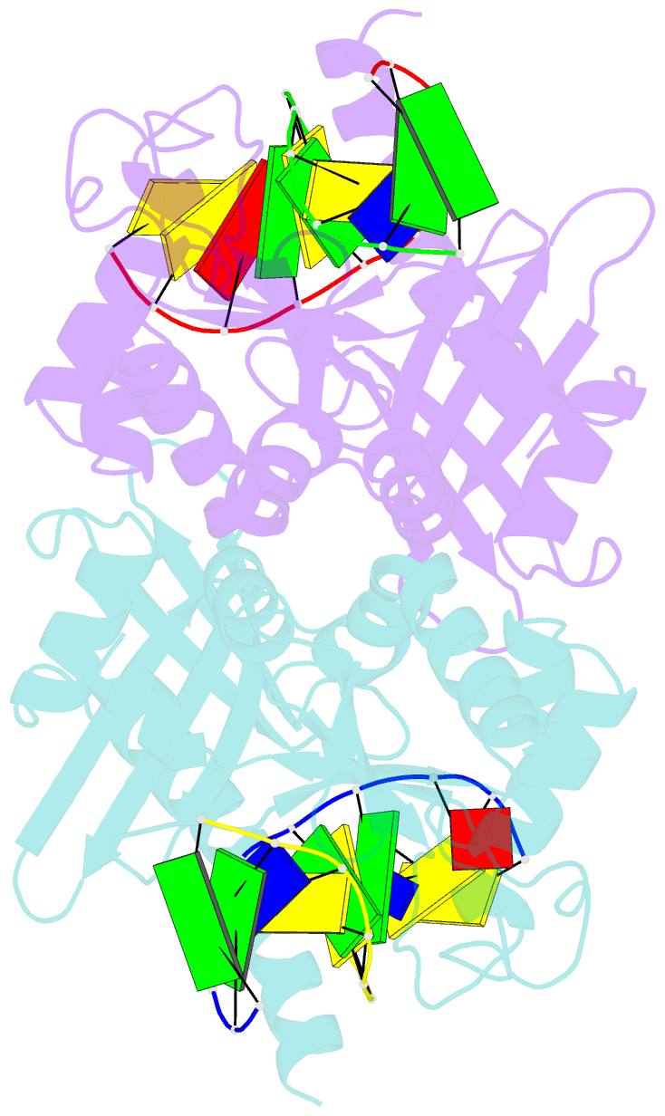
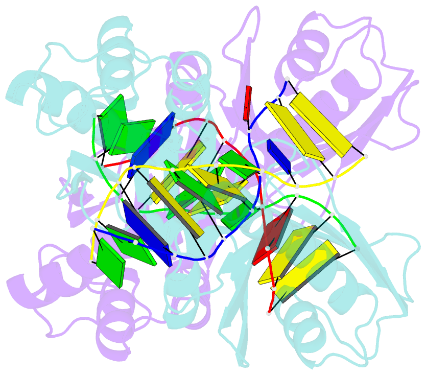
- Crystal form i of pre-reactive complex of restriction endonuclease hinp1i with cognate DNA and calcium ion
- Reference
- Horton JR, Zhang X, Maunus R, Yang Z, Wilson GG, Roberts RJ, Cheng X (2006): "DNA nicking by HinP1I endonuclease: bending, base flipping and minor groove expansion." Nucleic Acids Res., 34, 939-948. doi: 10.1093/nar/gkj484.
- Abstract
- HinP1I recognizes and cleaves the palindromic tetranucleotide sequence G downward arrowCGC in DNA. We report three structures of HinP1I-DNA complexes: in the presence of Ca(2+) (pre-reactive complex), in the absence of metal ion (binary complex) and in the presence of Mg(2+) (post-reactive complex). HinP1I forms a back-to-back dimer with two active sites and two DNA duplexes bound on the outer surfaces of the dimer facing away from each other. The 10 bp DNA duplexes undergo protein-induced distortions exhibiting features of A-, B- and Z-conformations: bending on one side (by intercalation of a phenylalanine side chain into the major groove), base flipping on the other side of the recognition site (by expanding the step rise distance of the local base pair to Z-form) and a local A-form conformation between the two central C:G base pairs of the recognition site (by binding of the N-terminal helix in the minor groove). In the pre- and post-reactive complexes, two metals (Ca(2+) or Mg(2+)) are found in the active site. The enzyme appears to cleave DNA sequentially, hydrolyzing first one DNA strand, as seen in the post-reactive complex in the crystalline state, and then the other, as supported by the observation that, in solution, a nicked DNA intermediate accumulates before linearization.