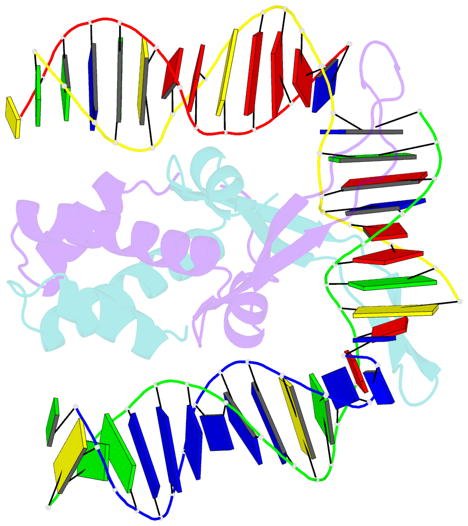
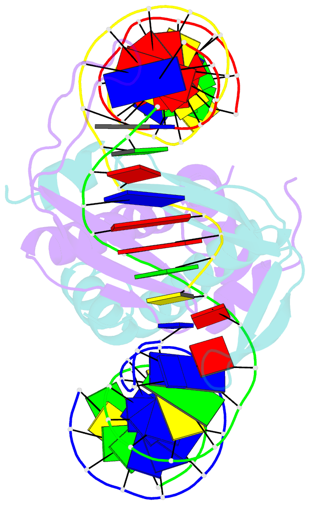
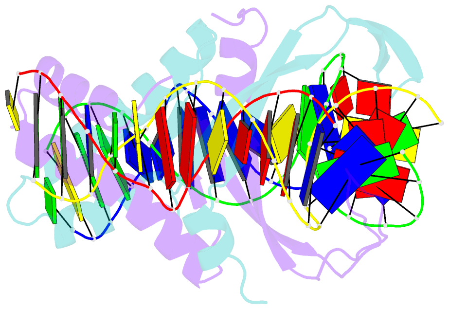
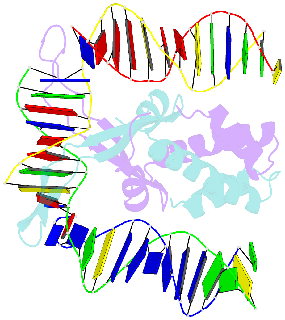
Summary information and primary citation
- PDB-id
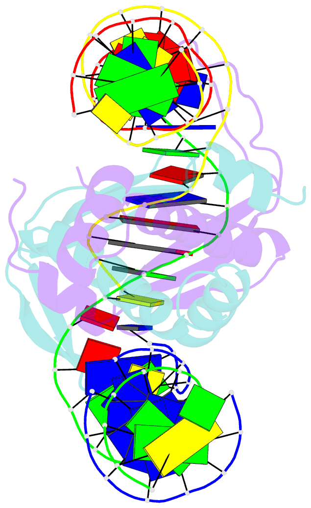
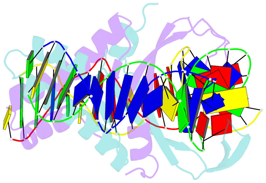
- 2ht0; SNAP-derived features in text and JSON formats;
DNAproDB
- Class
- transcription-DNA
- Method
- X-ray (2.0 Å)
- Summary
- Ihf bound to doubly nicked DNA
- Reference
- Swinger KK, Rice PA (2007): "Structure-based Analysis of HU-DNA Binding." J.Mol.Biol., 365, 1005-1016. doi: 10.1016/j.jmb.2006.10.024.
- Abstract
- HU and IHF are prokaryotic proteins that induce very large bends in DNA. They are present in high concentrations in the bacterial nucleoid and aid in chromosomal compaction. They also function as regulatory cofactors in many processes, such as site-specific recombination and the initiation of replication and transcription. HU and IHF have become paradigms for understanding DNA bending and indirect readout of sequence. While IHF shows significant sequence specificity, HU binds preferentially to certain damaged or distorted DNAs. However, none of the structurally diverse HU substrates previously studied in vitro is identical with the distorted substrates in the recently published Anabaena HU(AHU)-DNA cocrystal structures. Here, we report binding affinities for AHU and the DNA in the cocrystal structures. The binding free energies for formation of these AHU-DNA complexes range from approximately 10-14.5 kcal/mol, representing K(d) values in the nanomolar to low picomolar range, and a maximum stabilization of at least approximately 6.3 kcal/mol relative to complexes with undistorted, non-specific DNA. We investigated IHF binding and found that appropriate structural distortions can greatly enhance its affinity. On the basis of the coupling of structural and relevant binding data, we estimate the amount of conformational strain in an IHF-mediated DNA kink that is relieved by a nick (at least 0.76 kcal/mol) and pinpoint the location of the strain. We show that AHU has a sequence preference for an A+T-rich region in the center of its DNA-binding site, correlating with an unusually narrow minor groove. This is similar to sequence preferences shown by the eukaryotic nucleosome.