Summary information and primary citation
- PDB-id
- 2i82; SNAP-derived features in text and JSON formats;
DNAproDB
- Class
- lyase-RNA
- Method
- X-ray (2.05 Å)
- Summary
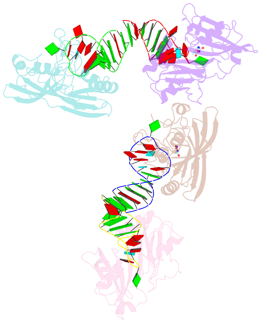
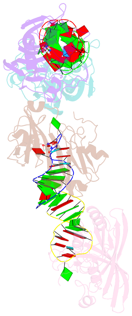
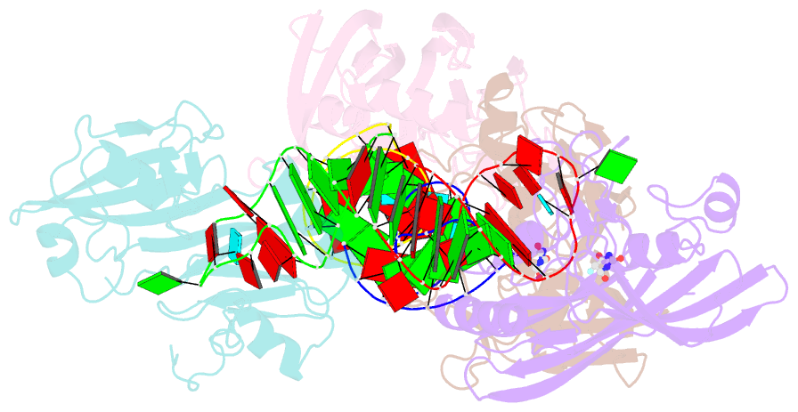
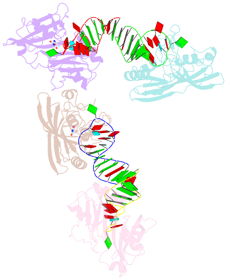
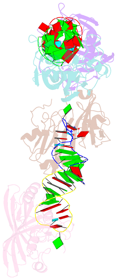
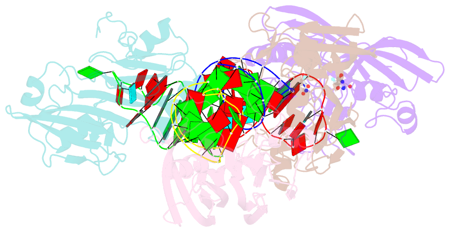
- Crystal structure of pseudouridine synthase rlua: indirect sequence readout through protein-induced RNA structure
- Reference
- Hoang C, Chen J, Vizthum CA, Kandel JM, Hamilton CS, Mueller EG, Ferre-D'Amare AR (2006): "Crystal structure of pseudouridine synthase RluA: indirect sequence readout through protein-induced RNA structure." Mol.Cell, 24, 535-545. doi: 10.1016/j.molcel.2006.09.017.
- Abstract
- RluA is a dual-specificity enzyme responsible for pseudouridylating 23S rRNA and several tRNAs. The 2.05 A resolution structure of RluA bound to a substrate RNA comprising the anticodon stem loop of tRNA(Phe) reveals that enzyme binding induces a dramatic reorganization of the RNA. Instead of adopting its canonical U turn conformation, the anticodon loop folds into a new structure with a reverse-Hoogsteen base pair and three flipped-out nucleotides. Sequence conservation, the cocrystal structure, and the results of structure-guided mutagenesis suggest that RluA recognizes its substrates indirectly by probing RNA loops for their ability to adopt the reorganized fold. The planar, cationic side chain of an arginine intercalates between the reverse-Hoogsteen base pair and the bottom pair of the anticodon stem, flipping the nucleotide to be modified into the active site of RluA. Sequence and structural comparisons suggest that pseudouridine synthases of the RluA, RsuA, and TruA families employ an equivalent arginine for base flipping.