Summary information and primary citation
- PDB-id
- 2kmk; SNAP-derived features in text and JSON formats;
DNAproDB
- Class
- DNA binding protein-DNA
- Method
- NMR
- Summary
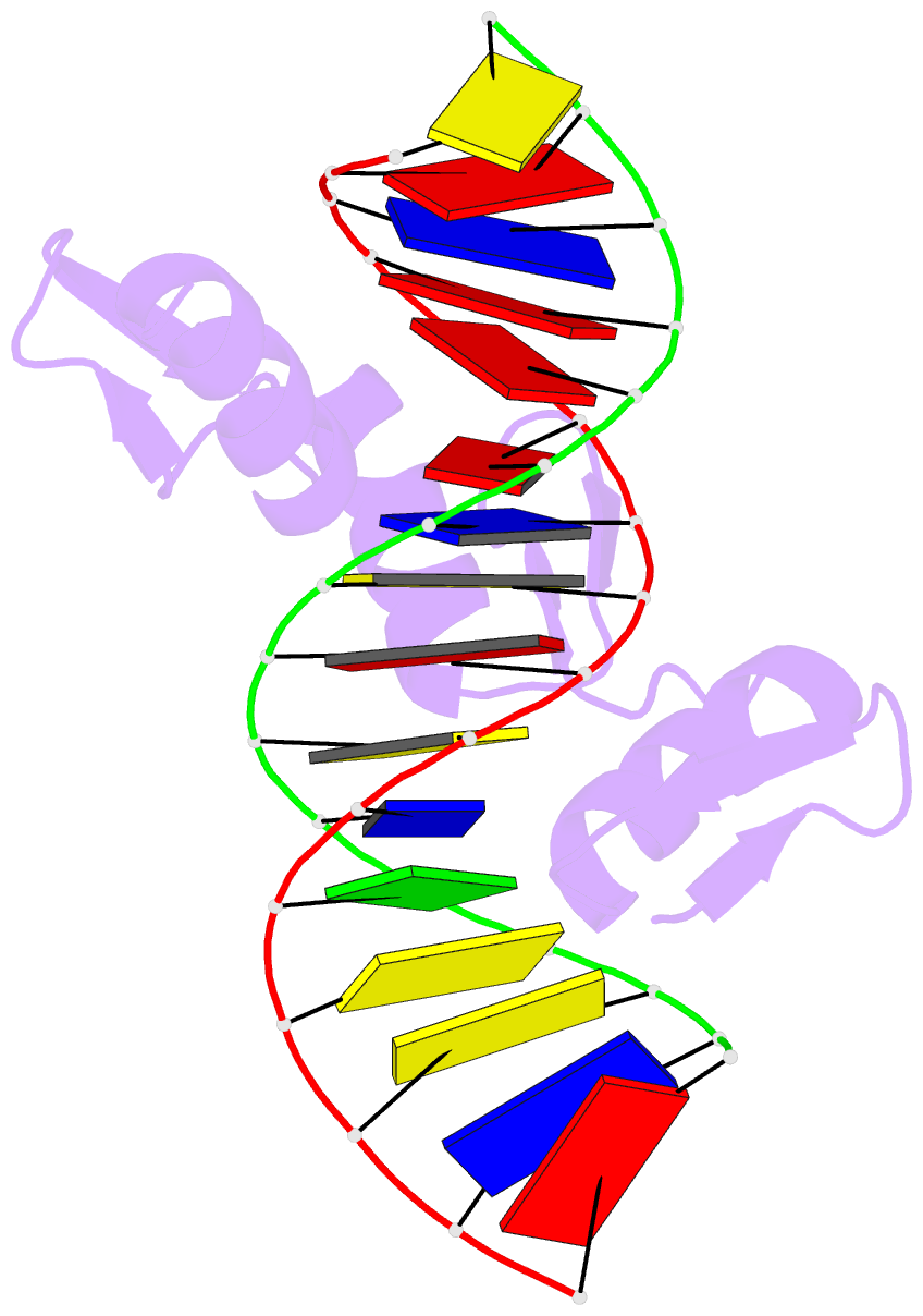
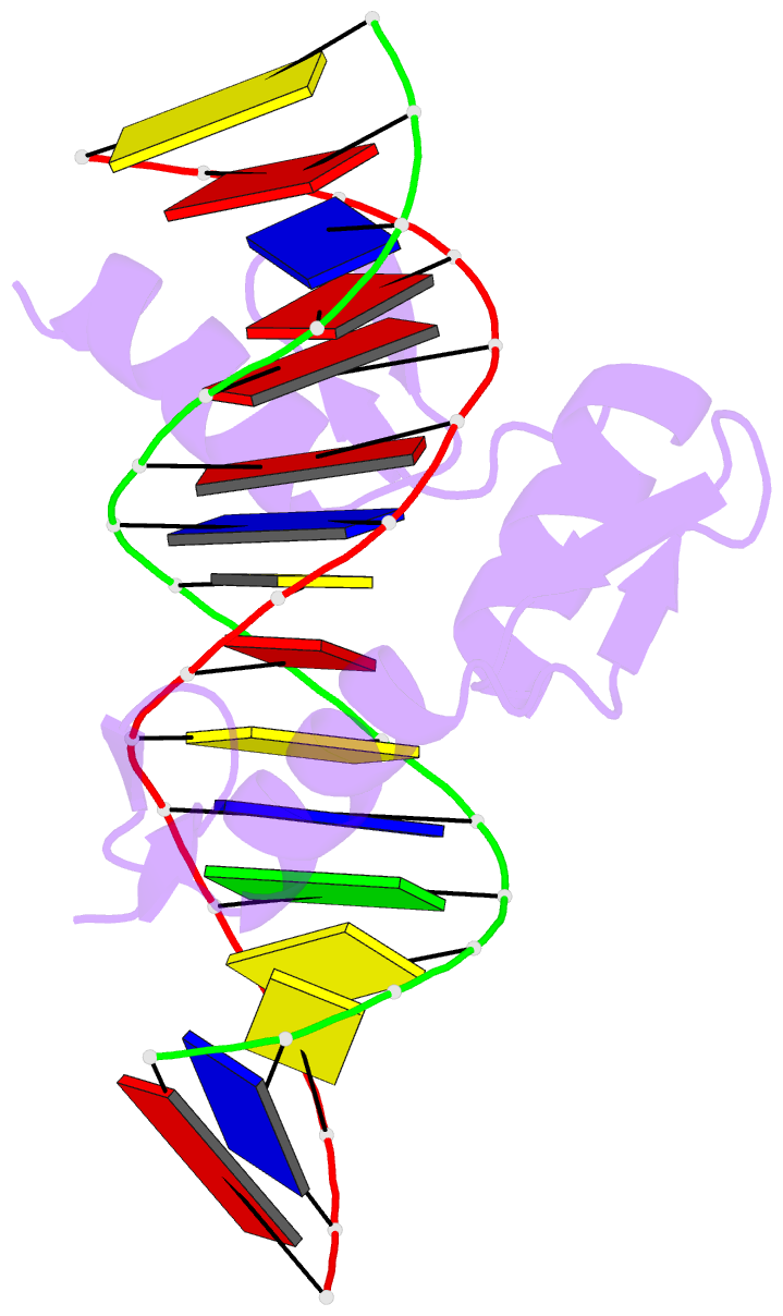
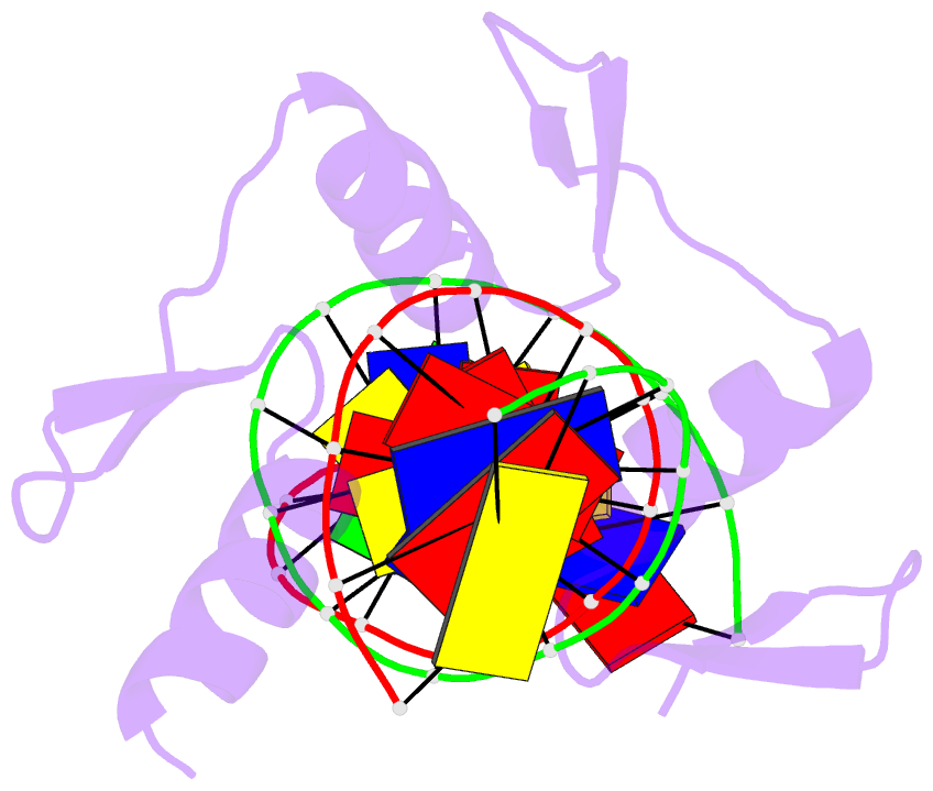
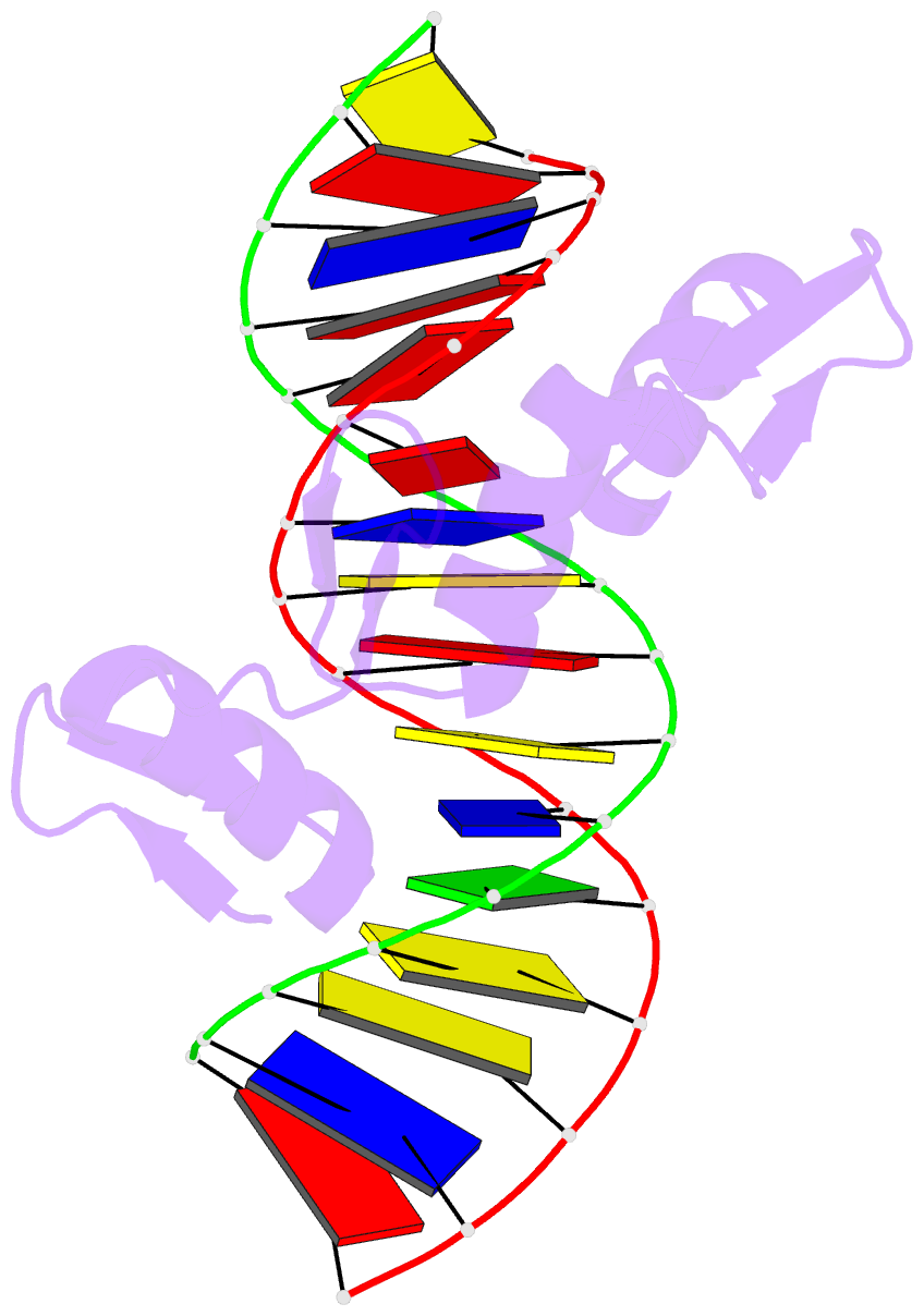
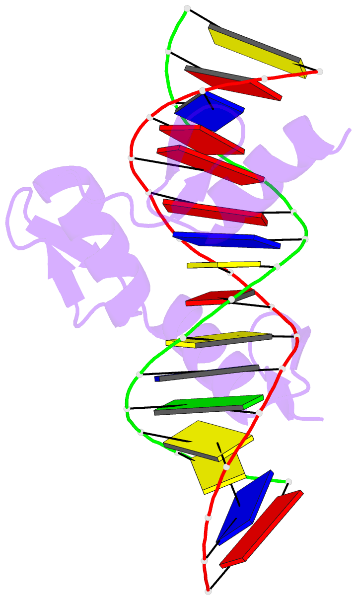
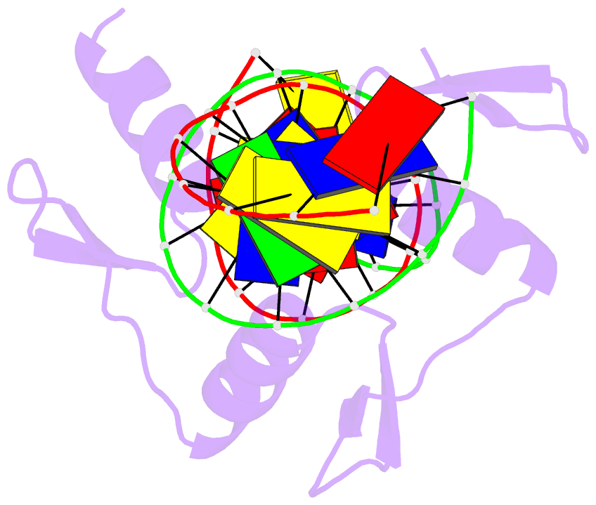
- Gfi-1 zinc fingers 3-5 complexed with DNA
- Reference
- Lee S, Doddapaneni K, Hogue A, McGhee L, Meyers S, Wu Z (2010): "Solution structure of Gfi-1 zinc domain bound to consensus DNA." J.Mol.Biol., 397, 1055-1066. doi: 10.1016/j.jmb.2010.02.006.
- Abstract
- Gfi-1 is a crucial transcriptional repressor for the precise regulation of cell proliferation and differentiation in hematopoiesis. Recently, this protein has also been demonstrated to be capable of restricting the proliferation of hematopoietic stem cells, a process that appears to be vital for the long-term competency of hematopoietic stem cells. These two seemingly opposite outcomes of regulation are likely to arise from its interactions with a variety of cellular partners. Such interactions can directly affect the genes that Gfi-1 recognizes through its DNA binding zinc-finger domain. In this work, we report the determination of the solution structure of Gfi-1 zinc fingers 3-5 in complex with a 16-mer consensus DNA using multidimensional NMR method. Unlike a proposed minor-groove binding model based on methylation interference experiments, our structure clearly shows that Gfi-1 zinc fingers 3-5 bind into the major groove of the target DNA reminiscent of canonical C(2)H(2) zinc-finger domains. The fourth and fifth zinc fingers recognize the AATC core sequence by forming base-specific hydrogen bonds between the side chains of Asn382, Gln379, and Asp354 and the bases of the invariant adenines and cytosine. Overall, the current work provides valuable insight into the structural determinants for DNA binding specificity, in particular for the TCA triplet that has not been observed in any other structures of zinc finger-DNA complexes, as well as molecular rationales for a naturally occurring mutation that causes acute myeloid leukemia.