Summary information and primary citation
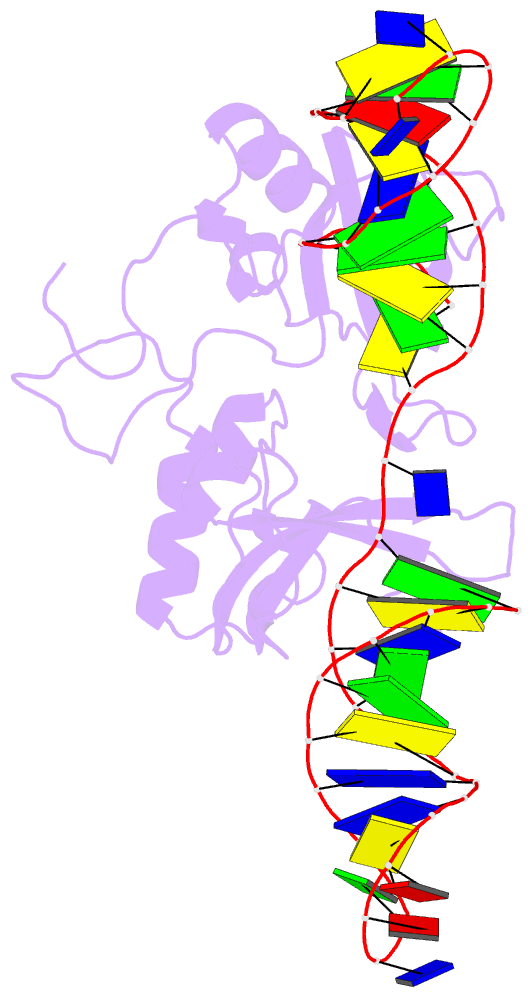
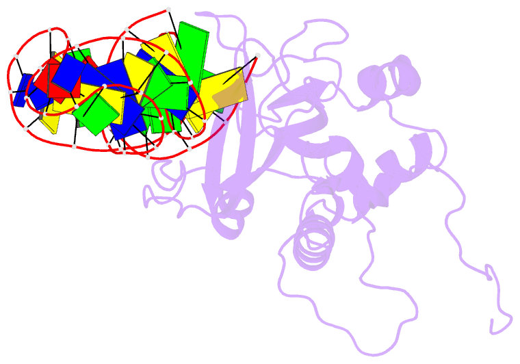
- PDB-id
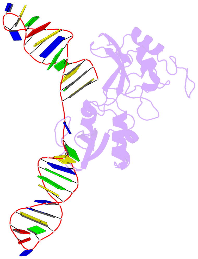
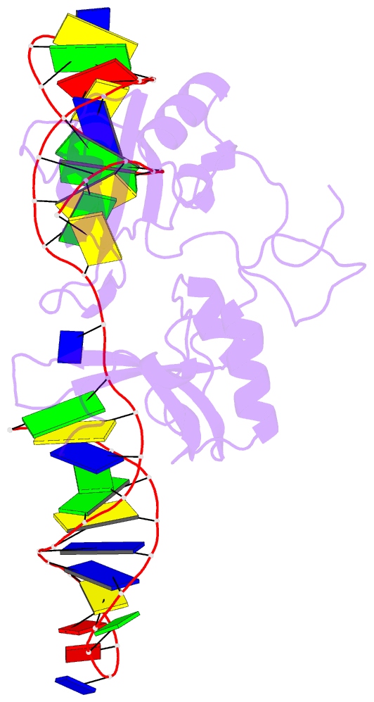
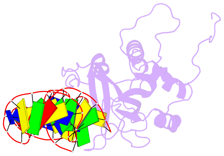
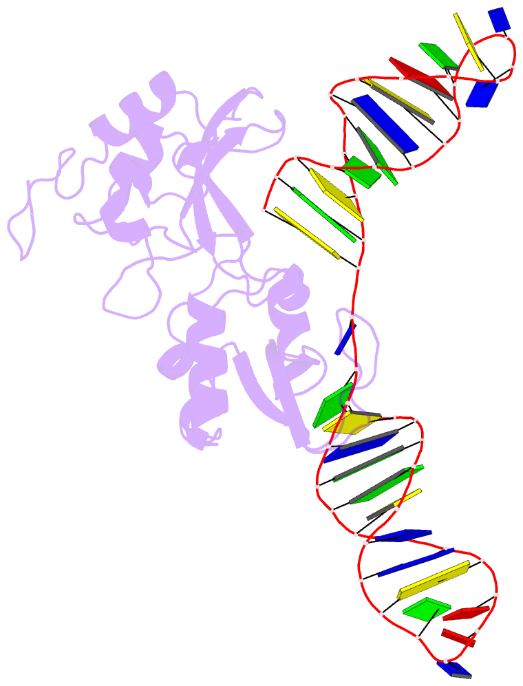
- 2n8a; SNAP-derived features in text and JSON formats;
DNAproDB
- Class
- transferase
- Method
- NMR
- Summary
- 1h, 13c and 15n chemical shift assignments and solution structure for parp-1 f1f2 domains in complex with a DNA single-strand break
- Reference
- Eustermann S, Wu WF, Langelier MF, Yang JC, Easton LE, Riccio AA, Pascal JM, Neuhaus D (2015): "Structural Basis of Detection and Signaling of DNA Single-Strand Breaks by Human PARP-1." Mol.Cell, 60, 742-754. doi: 10.1016/j.molcel.2015.10.032.
- Abstract
- Poly(ADP-ribose)polymerase 1 (PARP-1) is a key eukaryotic stress sensor that responds in seconds to DNA single-strand breaks (SSBs), the most frequent genomic damage. A burst of poly(ADP-ribose) synthesis initiates DNA damage response, whereas PARP-1 inhibition kills BRCA-deficient tumor cells selectively, providing the first anti-cancer therapy based on synthetic lethality. However, the mechanism underlying PARP-1's function remained obscure; inherent dynamics of SSBs and PARP-1's multi-domain architecture hindered structural studies. Here we reveal the structural basis of SSB detection and how multi-domain folding underlies the allosteric switch that determines PARP-1's signaling response. Two flexibly linked N-terminal zinc fingers recognize the extreme deformability of SSBs and drive co-operative, stepwise self-assembly of remaining PARP-1 domains to control the activity of the C-terminal catalytic domain. Automodification in cis explains the subsequent release of monomeric PARP-1 from DNA, allowing repair and replication to proceed. Our results provide a molecular framework for understanding PARP inhibitor action and, more generally, allosteric control of dynamic, multi-domain proteins.