Summary information and primary citation
- PDB-id
- 2vla; SNAP-derived features in text and JSON formats;
DNAproDB
- Class
- hydrolase
- Method
- X-ray (1.3 Å)
- Summary
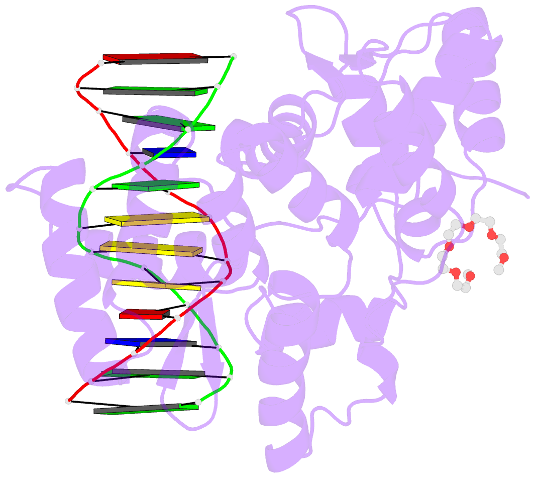
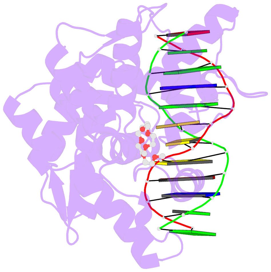
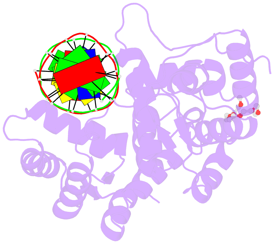
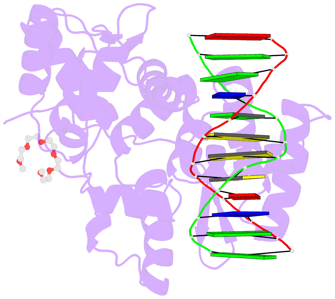
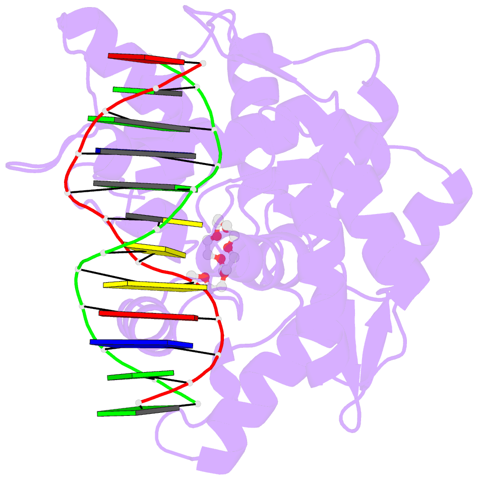
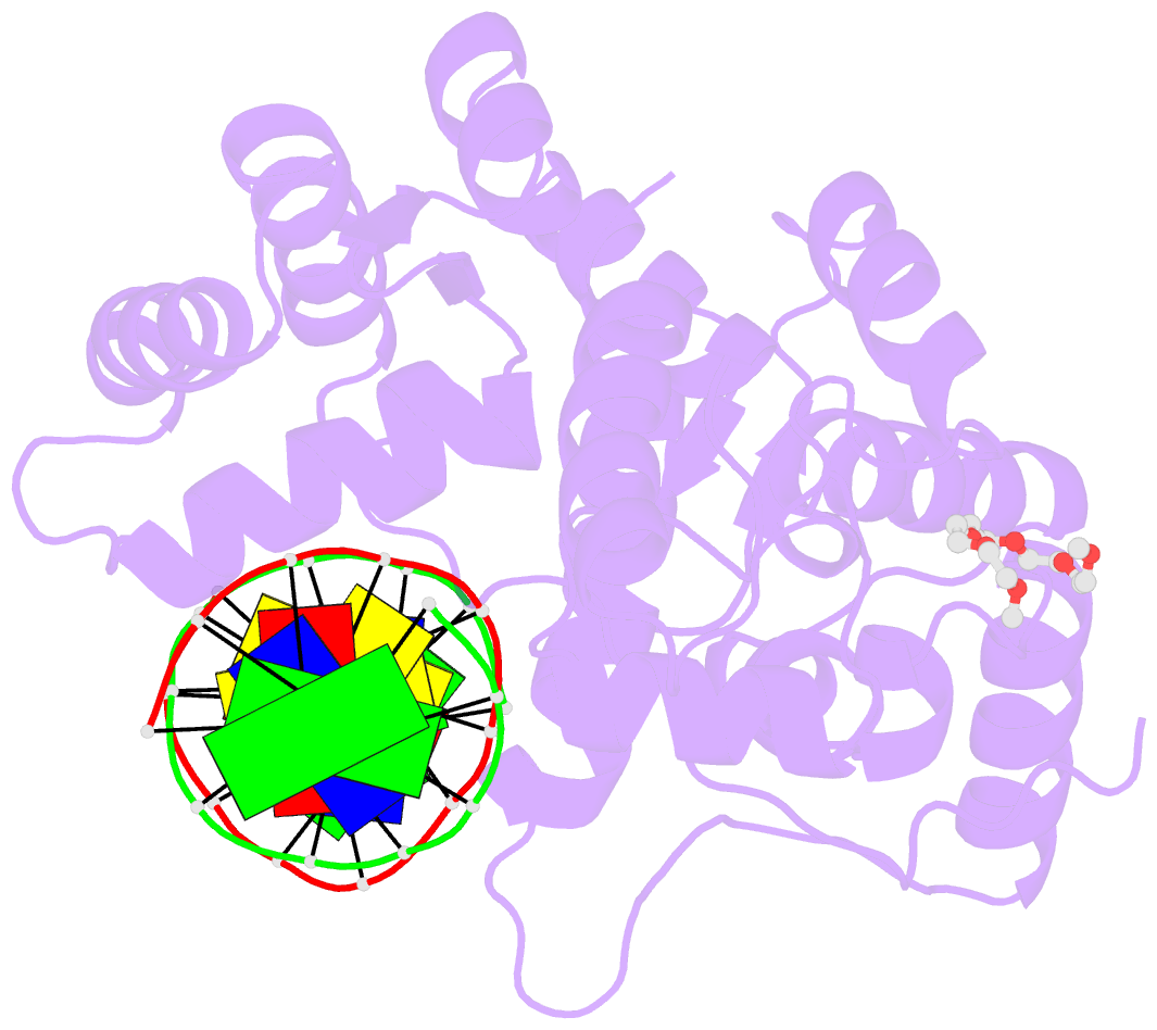
- Crystal structure of restriction endonuclease bpuji recognition domain in complex with cognate DNA
- Reference
- Sukackaite R, Grazulis S, Bochtler M, Siksnys V (2008): "The Recognition Domain of the Bpuji Restriction Endonuclease in Complex with Cognate DNA at 1.3-A Resolution." J.Mol.Biol., 378, 1084. doi: 10.1016/J.JMB.2008.03.041.
- Abstract
- Type IIS restriction endonucleases recognize asymmetric DNA sequences and cleave both DNA strands at fixed positions downstream of the recognition site. The restriction endonuclease BpuJI recognizes the asymmetric sequence 5'-CCCGT; however, it cuts at multiple sites in the vicinity of the target sequence. BpuJI consists of two physically separate domains, with catalytic and dimerization functions in the C-terminal domain and DNA recognition functions in the N-terminal domain. Here we report the crystal structure of the BpuJI recognition domain bound to cognate DNA at 1.3-A resolution. This region folds into two winged-helix subdomains, D1 and D2, interspaced by the DL subdomain. The D1 and D2 subdomains of BpuJI share structural similarity with the similar subdomains of the FokI DNA-binding domain; however, their orientations in protein-DNA complexes are different. Recognition of the 5'-CCCGT target sequence is achieved by BpuJI through the major groove contacts of amino acid residues located on both the helix-turn-helix motifs and the N-terminal arm. The role of these interactions in DNA recognition is also corroborated by mutational analysis.