Summary information and primary citation
- PDB-id
- 2xro; SNAP-derived features in text and JSON formats;
DNAproDB
- Class
- DNA-binding protein-DNA
- Method
- X-ray (3.4 Å)
- Summary
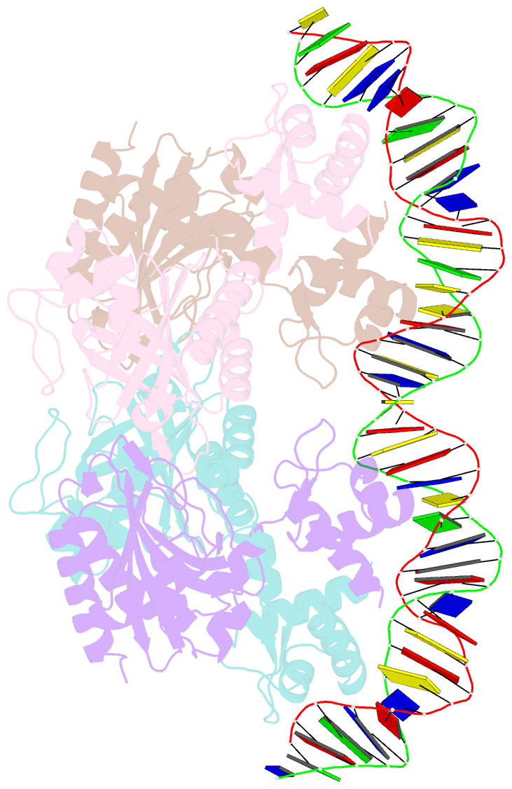
- Crystal structure of ttgv in complex with its DNA operator
- Reference
- Lu D, Fillet S, Meng C, Alguel Y, Kloppsteck P, Bergeron J, Krell T, Gallegos MT, Ramos J, Zhang X (2010): "Crystal Structure of Ttgv in Complex with its DNA Operator Reveals a General Model for Cooperative DNA Binding of Tetrameric Gene Regulators." Genes Dev., 24, 2556. doi: 10.1101/GAD.603510.
- Abstract
- The majority of bacterial gene regulators bind as symmetric dimers to palindromic DNA operators of 12-20 base pairs (bp). Multimeric forms of proteins, including tetramers, are able to recognize longer operator sequences in a cooperative manner, although how this is achieved is not well understood due to the lack of complete structural information. Models, instead of structures, of complete tetrameric assembly on DNA exist in literature. Here we present the crystal structures of the multidrug-binding protein TtgV, a gene repressor that controls efflux pumps, alone and in complex with a 42-bp DNA operator containing two TtgV recognition sites at 2.9 Å and 3.4 Å resolution. These structures represent the first full-length functional tetrameric protein in complex with its intact DNA operator containing two continuous recognition sites. TtgV binds to its DNA operator as a highly asymmetric tetramer and induces considerable distortions in the DNA, resulting in a 60° bend. Upon binding to its operator, TtgV undergoes large conformational changes at the monomeric, dimeric, and tetrameric levels. The structures here reveal a general model for cooperative DNA binding of tetrameric gene regulators and provide a structural basis for a large body of biochemical data and a reinterpretation of previous models for tetrameric gene regulators derived from partial structural data.


