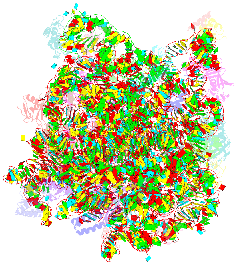
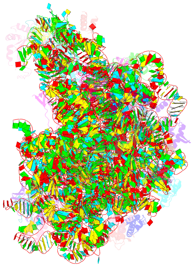
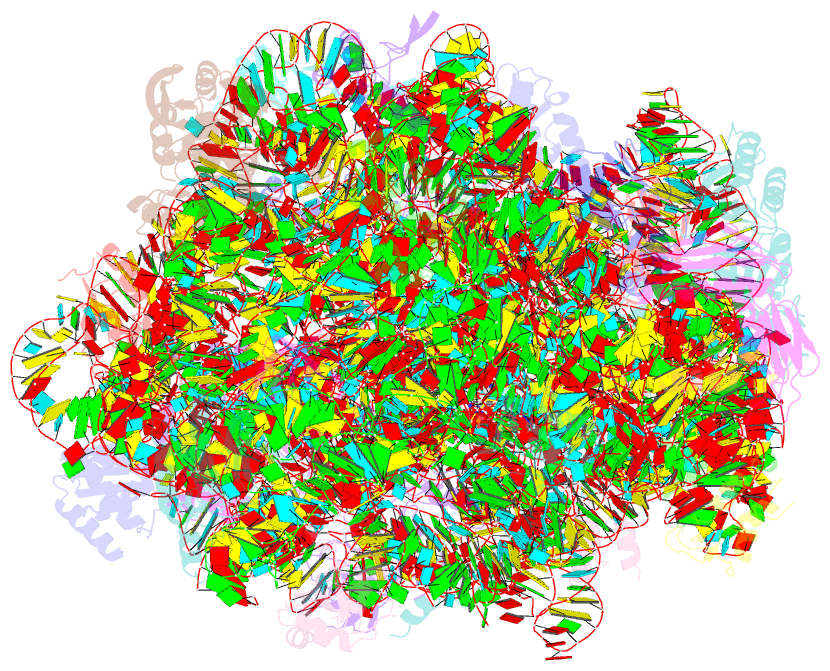
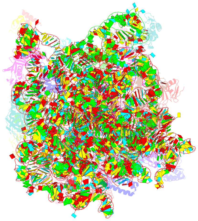
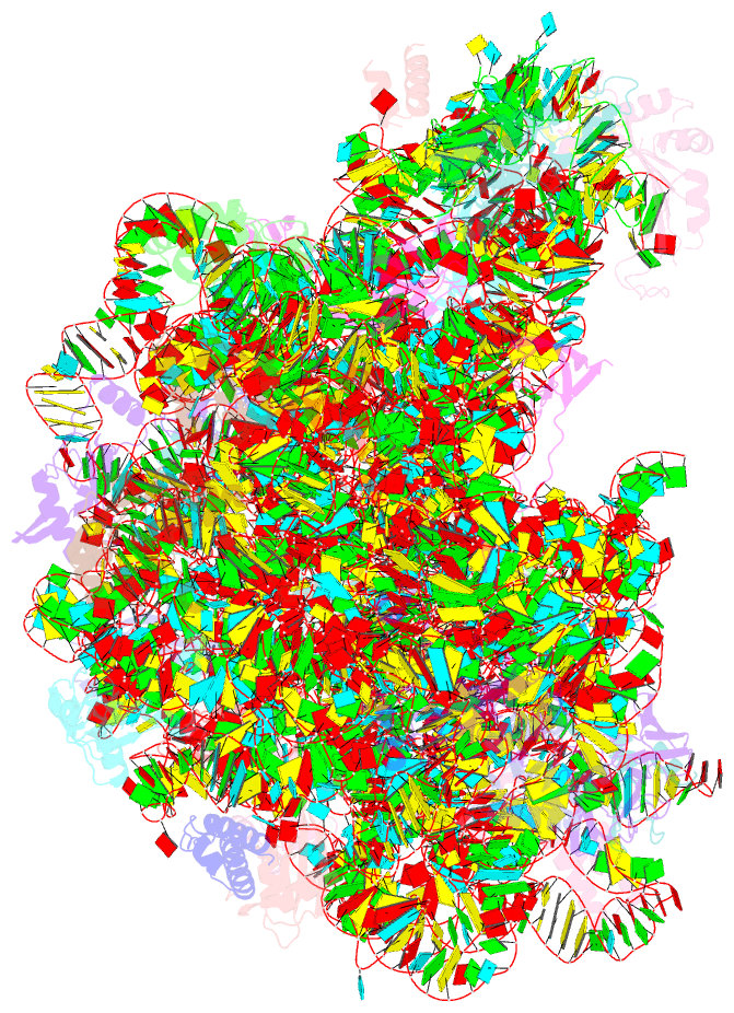
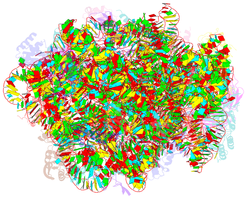
Summary information and primary citation
- PDB-id
- 3cme; SNAP-derived features in text and JSON formats;
DNAproDB
- Class
- ribosome
- Method
- X-ray (2.95 Å)
- Summary
- The structure of ca and cca-phe-cap-bio bound to the large ribosomal subunit of haloarcula marismortui
- Reference
- Simonovic M, Steitz TA (2008): "Peptidyl-CCA deacylation on the ribosome promoted by induced fit and the O3'-hydroxyl group of A76 of the unacylated A-site tRNA." Rna, 14, 2372-2378. doi: 10.1261/rna.1118908.
- Abstract
- The last step in ribosome-catalyzed protein synthesis is the hydrolytic release of the newly formed polypeptide from the P-site bound tRNA. Hydrolysis of the ester link of the peptidyl-tRNA is stimulated normally by the binding of release factors (RFs). However, an unacylated tRNA or just CCA binding to the ribosomal A site can also stimulate deacylation under some nonphysiological conditions. Although the sequence of events is well described by biochemical studies, the structural basis of the mechanism underlying this process is not well understood. Two new structures of the large ribosomal subunit of Haloarcula marismortui complexed with a peptidyl-tRNA analog in the P site and two oligonucleotide mimics of unacylated tRNA, CCA and CA, in the A site show that the binding of either CA or CCA induces a very similar conformational change in the peptidyl-transferase center as induced by aminoacyl-CCA. However, only CCA positions a water molecule appropriately to attack the carbonyl carbon of the peptidyl-tRNA and stabilizes the proper orientation of the ester link for hydrolysis. We, thus, conclude that both the ability of the O3'-hydroxyl group of the A-site A76 to position the water and the A-site CCA induced conformational change of the PTC are critical for the catalysis of the deacylation of the peptidyl-tRNA by CCA, and perhaps, an analogous mechanism is used by RFs.