Summary information and primary citation
- PDB-id
- 3fdq; SNAP-derived features in text and JSON formats;
DNAproDB
- Class
- DNA binding protein-DNA
- Method
- X-ray (1.75 Å)
- Summary
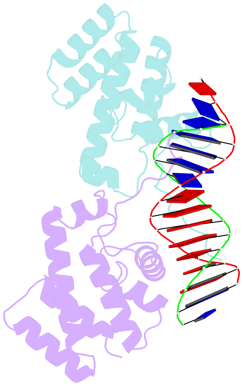
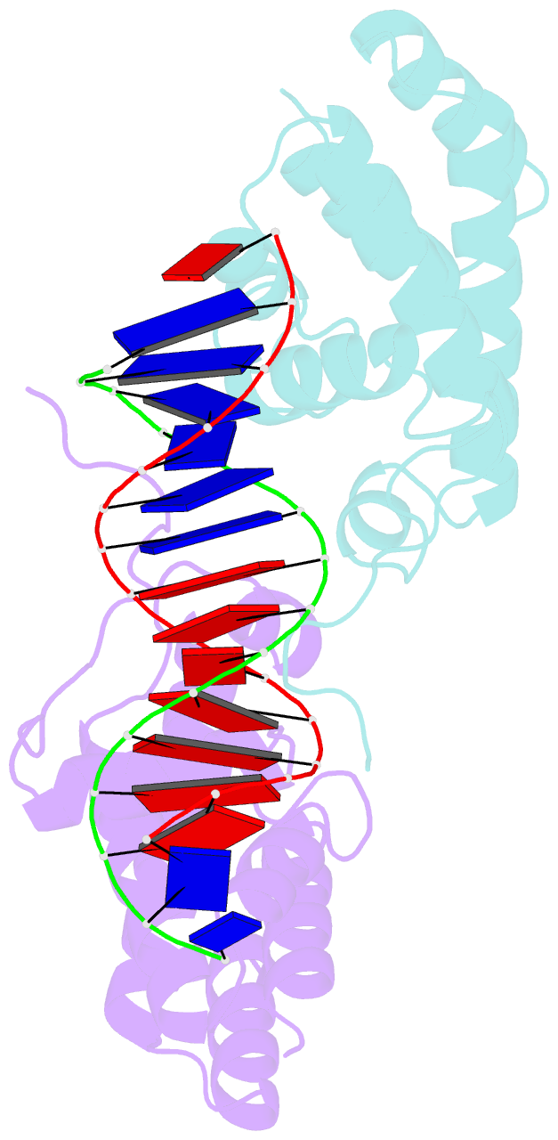
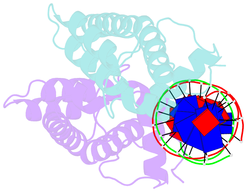
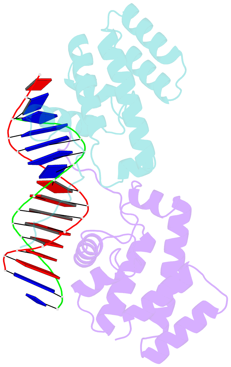
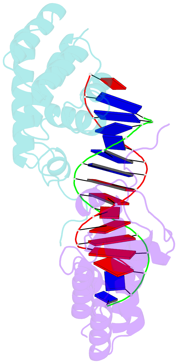
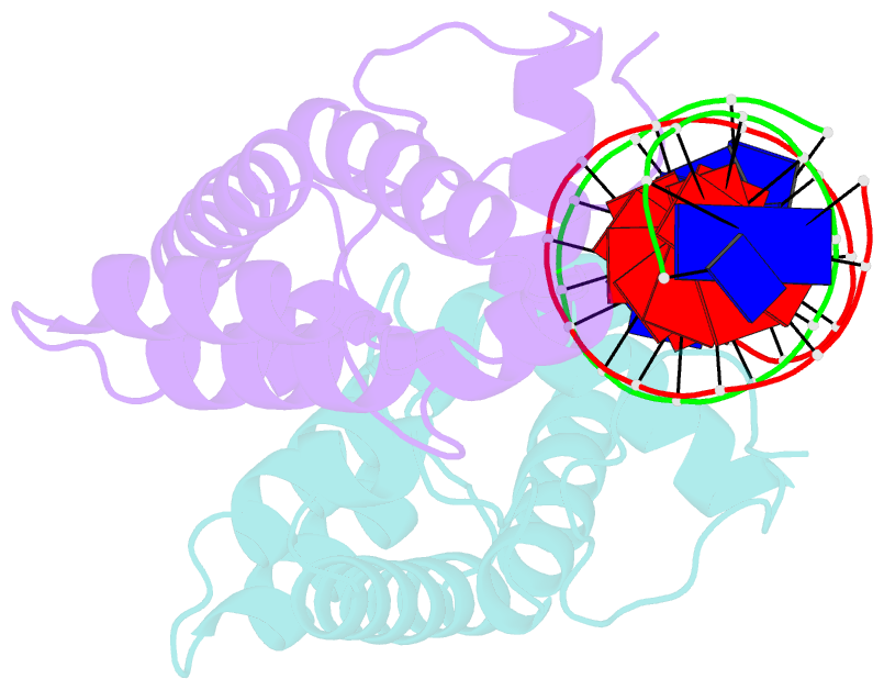
- Recognition of at-rich DNA binding sites by the mogr repressor
- Reference
- Shen A, Higgins DE, Panne D (2009): "Recognition of AT-Rich DNA Binding Sites by the MogR Repressor." Structure, 17, 769-777. doi: 10.1016/j.str.2009.02.018.
- Abstract
- The MogR transcriptional repressor of the intracellular pathogen Listeria monocytogenes recognizes AT-rich binding sites in promoters of flagellar genes to downregulate flagellar gene expression during infection. We describe here the 1.8 A resolution crystal structure of MogR bound to the recognition sequence 5' ATTTTTTAAAAAAAT 3' present within the flaA promoter region. Our structure shows that MogR binds as a dimer. Each half-site is recognized in the major groove by a helix-turn-helix motif and in the minor groove by a loop from the symmetry-related molecule, resulting in a "crossover" binding mode. This oversampling through minor groove interactions is important for specificity. The MogR binding site has structural features of A-tract DNA and is bent by approximately 52 degrees away from the dimer. The structure explains how MogR achieves binding specificity in the AT-rich genome of L. monocytogenes and explains the evolutionary conservation of A-tract sequence elements within promoter regions of MogR-regulated flagellar genes.