Summary information and primary citation
- PDB-id
- 3hqg; SNAP-derived features in text and JSON formats;
DNAproDB
- Class
- hydrolase-DNA
- Method
- X-ray (2.6 Å)
- Summary
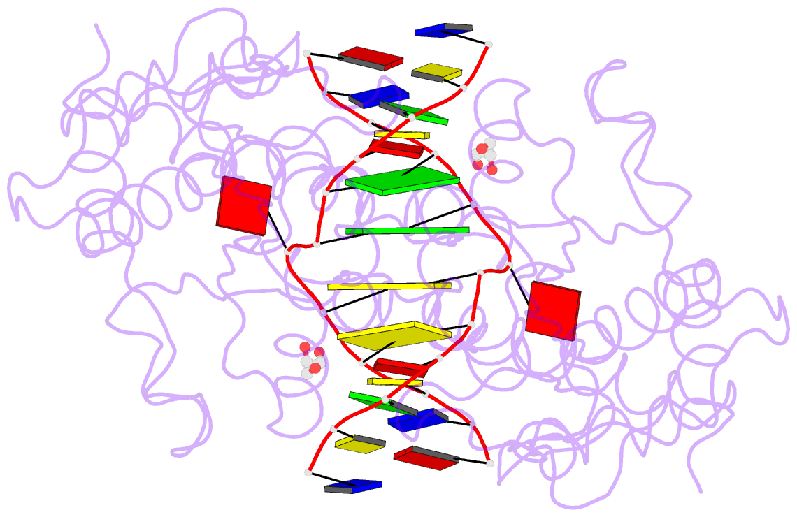
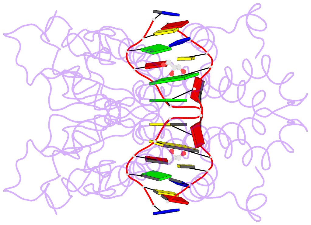
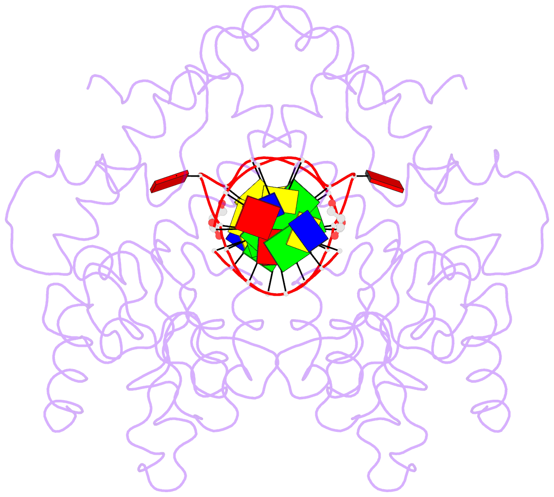
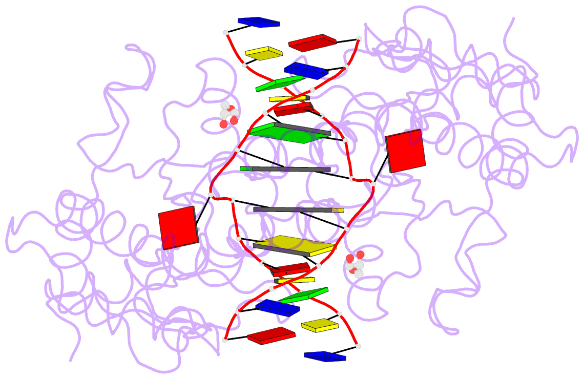
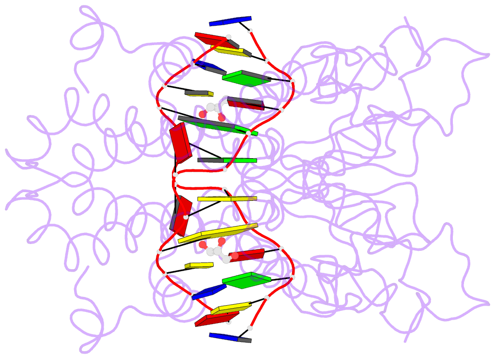
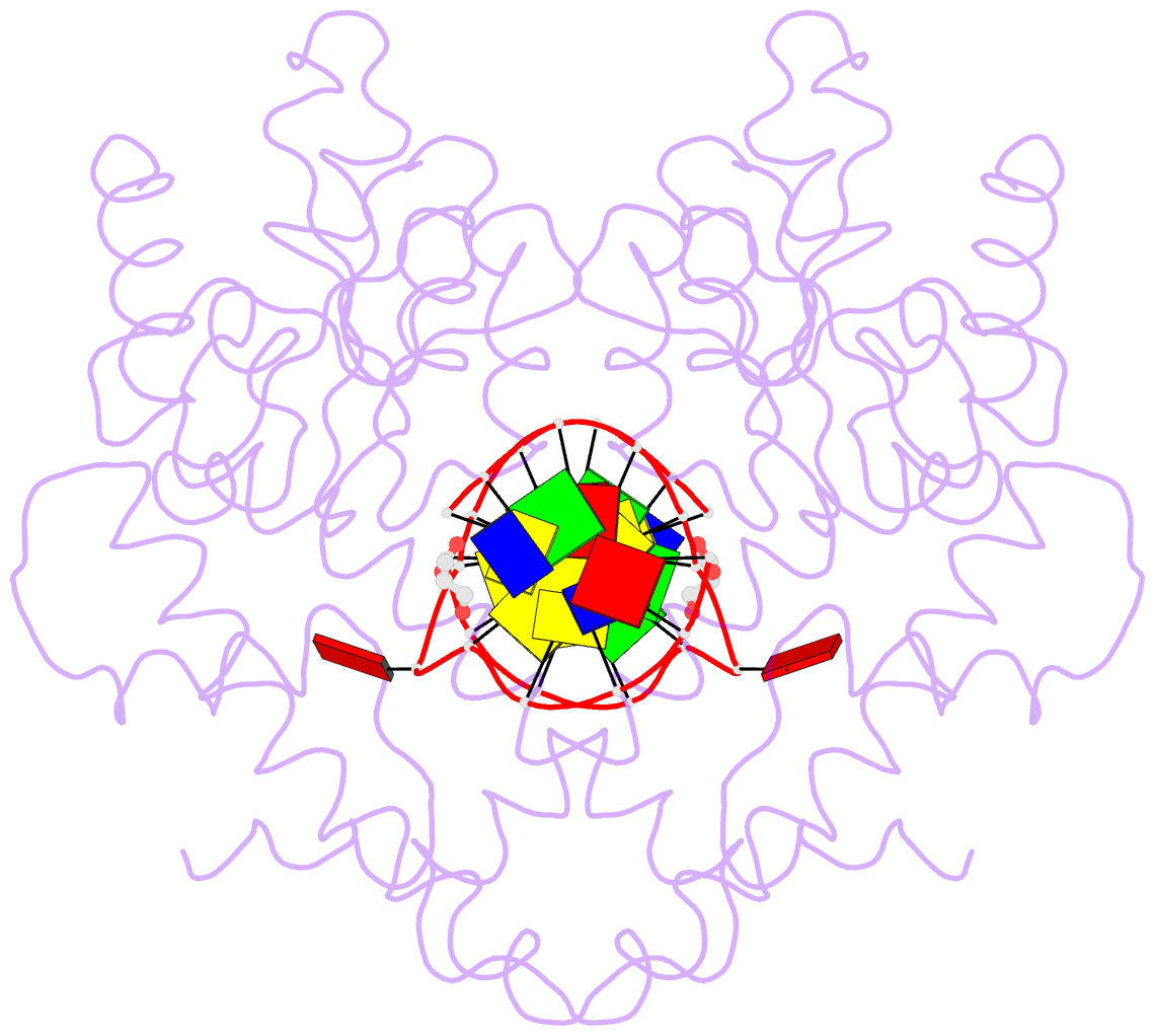
- Crystal structure of restriction endonuclease ecorii catalytic c-terminal domain in complex with cognate DNA
- Reference
- Golovenko D, Manakova E, Tamulaitiene G, Grazulis S, Siksnys V (2009): "Structural mechanisms for the 5'-CCWGG sequence recognition by the N- and C-terminal domains of EcoRII." Nucleic Acids Res., 37, 6613-6624. doi: 10.1093/nar/gkp699.
- Abstract
- EcoRII restriction endonuclease is specific for the 5'-CCWGG sequence (W stands for A or T); however, it shows no activity on a single recognition site. To activate cleavage it requires binding of an additional target site as an allosteric effector. EcoRII dimer consists of three structural units: a central catalytic core, made from two copies of the C-terminal domain (EcoRII-C), and two N-terminal effector DNA binding domains (EcoRII-N). Here, we report DNA-bound EcoRII-N and EcoRII-C structures, which show that EcoRII combines two radically different structural mechanisms to interact with the effector and substrate DNA. The catalytic EcoRII-C dimer flips out the central T:A base pair and makes symmetric interactions with the CC:GG half-sites. The EcoRII-N effector domain monomer binds to the target site asymmetrically in a single defined orientation which is determined by specific hydrogen bonding and van der Waals interactions with the central T:A pair in the major groove. The EcoRII-N mode of the target site recognition is shared by the large class of higher plant transcription factors of the B3 superfamily.