Summary information and primary citation
- PDB-id
- 3id5; SNAP-derived features in text and JSON formats;
DNAproDB
- Class
- transferase-ribosomal protein-RNA
- Method
- X-ray (4.01 Å)
- Summary
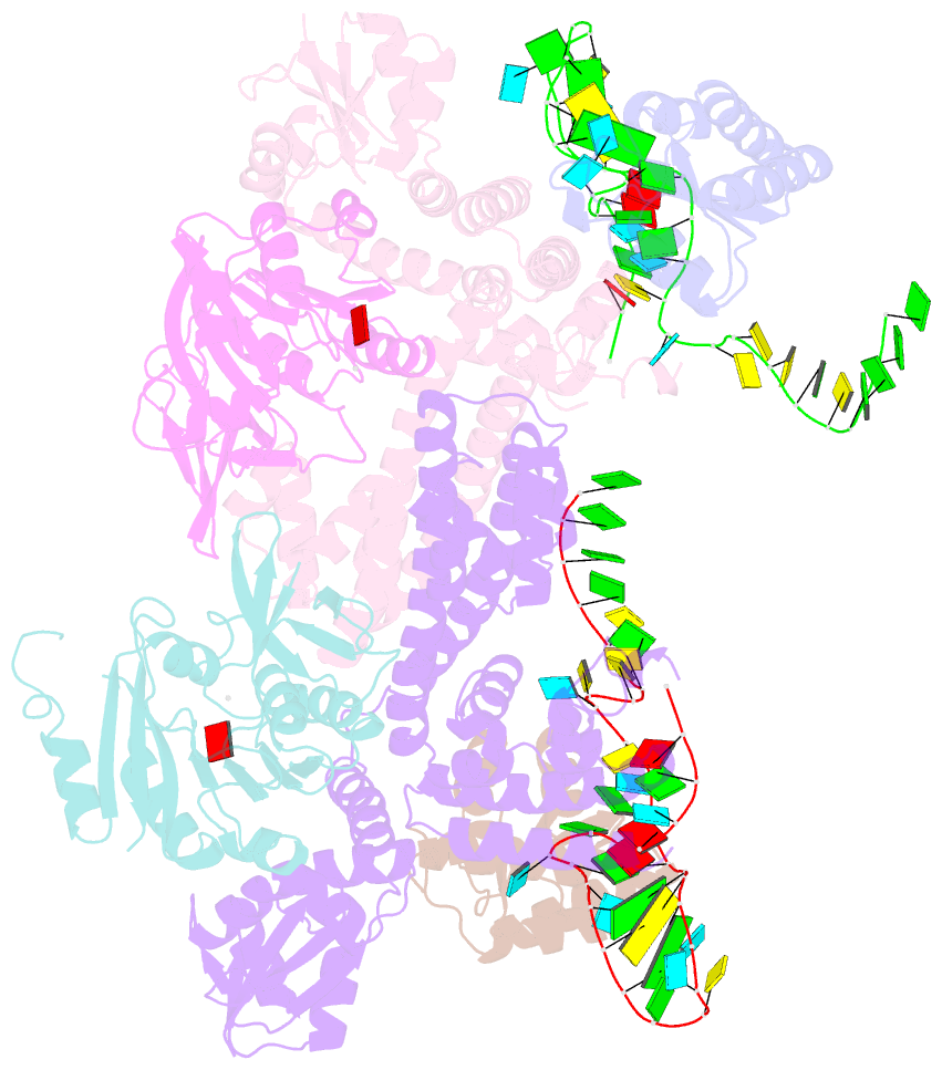
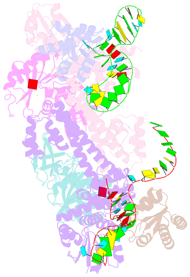
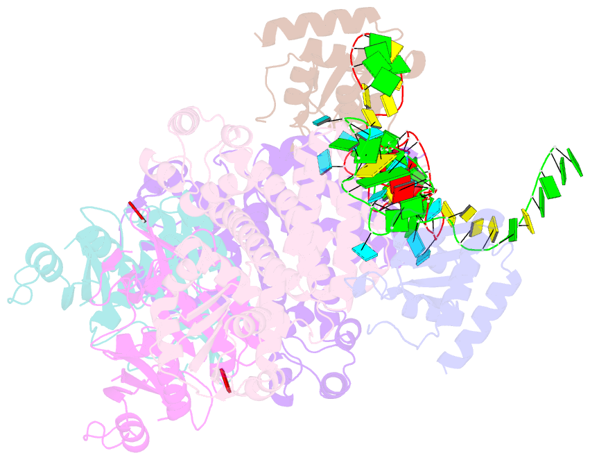
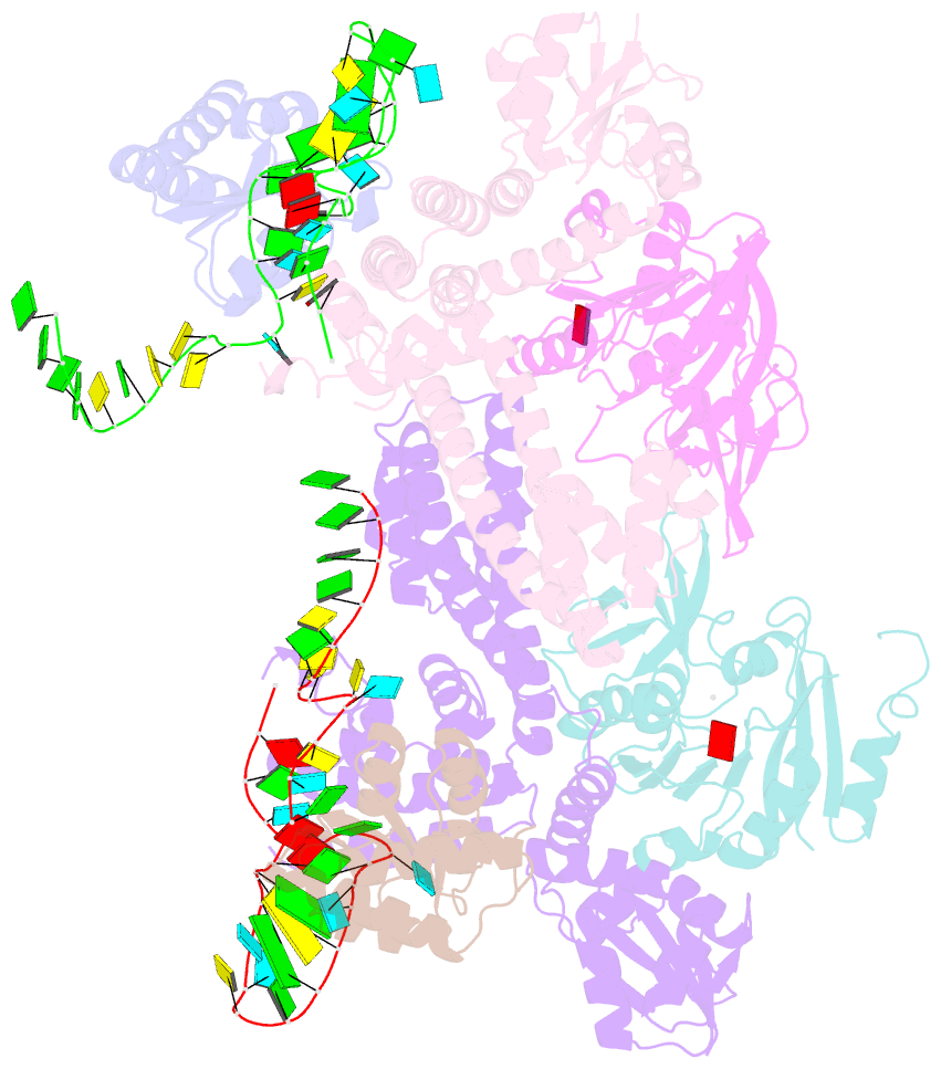
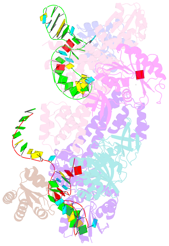
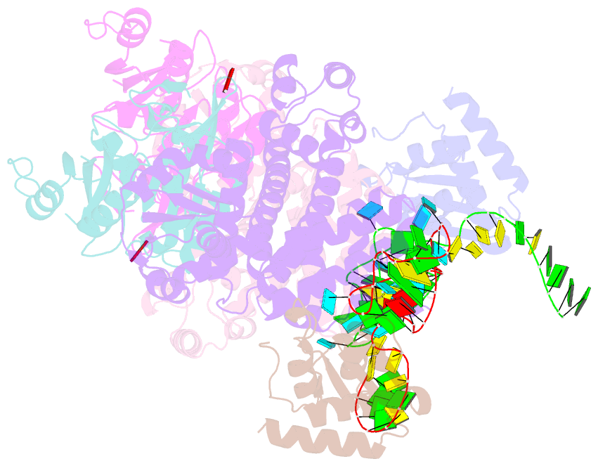
- Crystal structure of sulfolobus solfataricus c-d rnp assembled with nop5, fibrillarin, l7ae and a split half c-d RNA
- Reference
- Ye K, Jia R, Lin J, Ju M, Peng J, Xu A, Zhang L (2009): "Structural organization of box C/D RNA-guided RNA methyltransferase." Proc.Natl.Acad.Sci.USA, 106, 13808-13813. doi: 10.1073/pnas.0905128106.
- Abstract
- Box C/D guide RNAs are abundant noncoding RNAs that primarily function to direct the 2'-O-methylation of specific nucleotides by base-pairing with substrate RNAs. In archaea, a bipartite C/D RNA assembles with L7Ae, Nop5, and the methyltransferase fibrillarin into a modification enzyme with unique substrate specificity. Here, we determined the crystal structure of an archaeal C/D RNA-protein complex (RNP) composed of all 3 core proteins and an engineered half-guide RNA at 4 A resolution, as well as 2 protein substructures at higher resolution. The RNP structure reveals that the C-terminal domains of Nop5 in the dimeric complex provide symmetric anchoring sites for 2 L7Ae-associated kink-turn motifs of the C/D RNA. A prominent protrusion in Nop5 seems to be important for guide RNA organization and function and for discriminating the structurally related U4 snRNA. Multiple conformations of the N-terminal domain of Nop5 and its associated fibrillarin in different structures indicate the inherent flexibility of the catalytic module, suggesting that a swinging motion of the catalytic module is part of the enzyme mechanism. We also built a model of a native C/D RNP with substrate and fibrillarin in an active conformation. Our results provide insight into the overall organization and mechanism of action of C/D RNA-guided RNA methyltransferases.