Summary information and primary citation
- PDB-id
- 3irr; SNAP-derived features in text and JSON formats;
DNAproDB
- Class
- hydrolase-DNA
- Method
- X-ray (2.65 Å)
- Summary
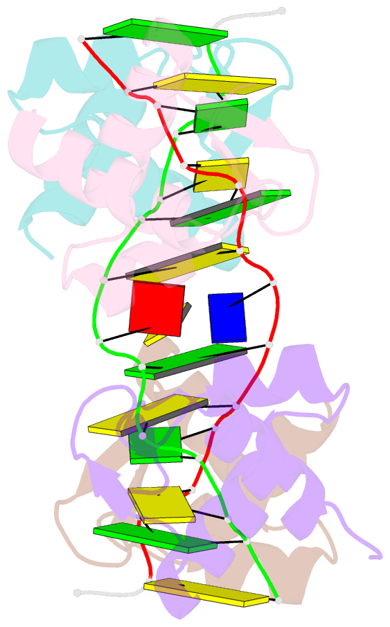
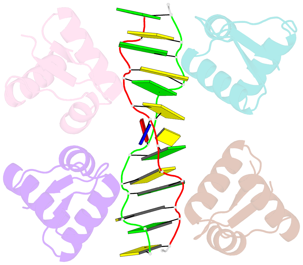
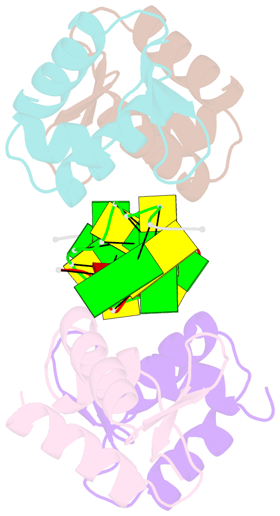
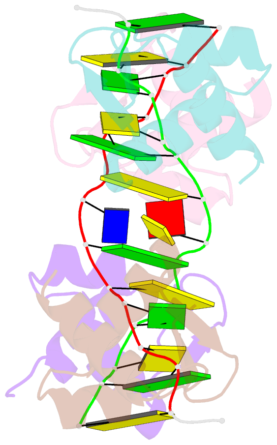
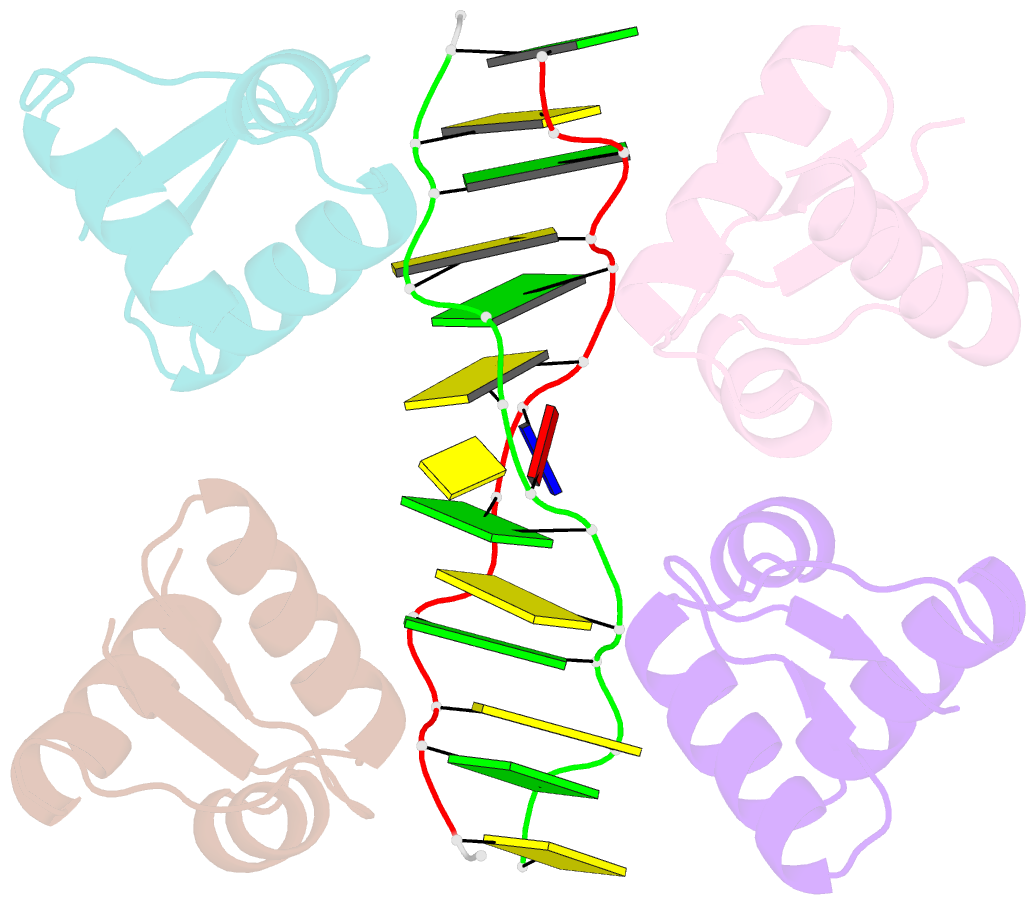
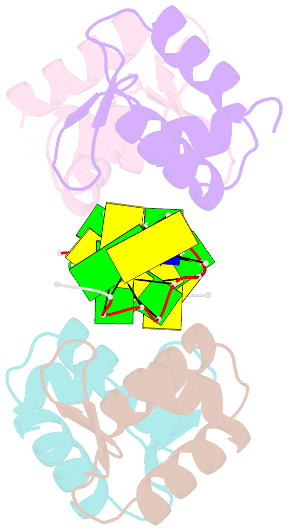
- Crystal structure of a z-z junction (with hepes intercalating)
- Reference
- de Rosa M, de Sanctis D, Rosario AL, Archer M, Rich A, Athanasiadis A, Carrondo MA (2010): "Crystal structure of a junction between two Z-DNA helices." Proc.Natl.Acad.Sci.USA, 107, 9088-9092. doi: 10.1073/pnas.1003182107.
- Abstract
- The double helix of DNA, when composed of dinucleotide purine-pyrimidine repeats, can adopt a left-handed helical structure called Z-DNA. For reasons not entirely understood, such dinucleotide repeats in genomic sequences have been associated with genomic instability leading to cancer. Adoption of the left-handed conformation results in the formation of conformational junctions: A B-to-Z junction is formed at the boundaries of the helix, whereas a Z-to-Z junction is commonly formed in sequences where the dinucleotide repeat is interrupted by single base insertions or deletions that bring neighboring helices out of phase. B-Z junctions are shown to result in exposed nucleotides vulnerable to chemical or enzymatic modification. Here we describe the three-dimensional structure of a Z-Z junction stabilized by Zalpha, the Z-DNA binding domain of the RNA editing enzyme ADAR1. We show that the junction structure consists of a single base pair and leads to partial or full disruption of the helical stacking. The junction region allows intercalating agents to insert themselves into the left-handed helix, which is otherwise resistant to intercalation. However, unlike a B-Z junction, in this structure the bases are not fully extruded, and the stacking between the two left-handed helices is not continuous.