Summary information and primary citation
- PDB-id
- 3qeb; SNAP-derived features in text and JSON formats;
DNAproDB
- Class
- hydrolase-DNA
- Method
- X-ray (3.0 Å)
- Summary
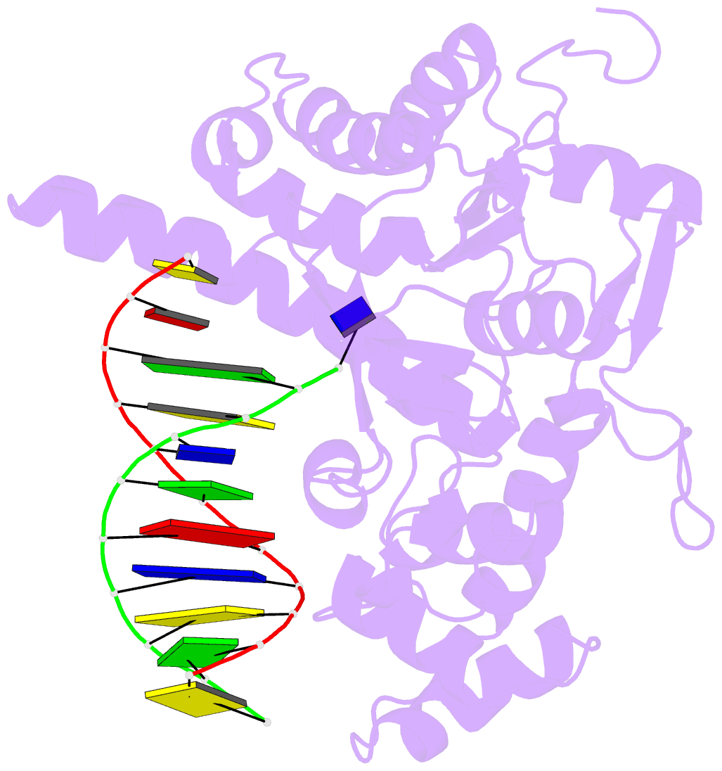
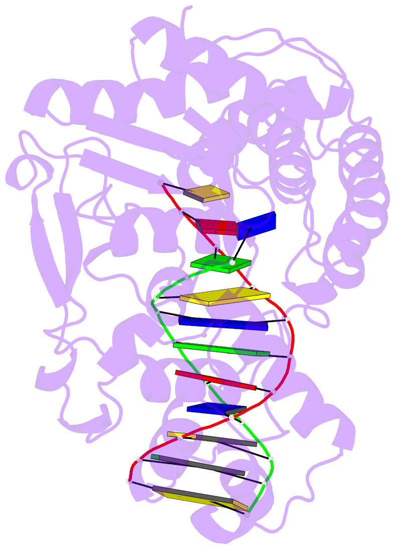
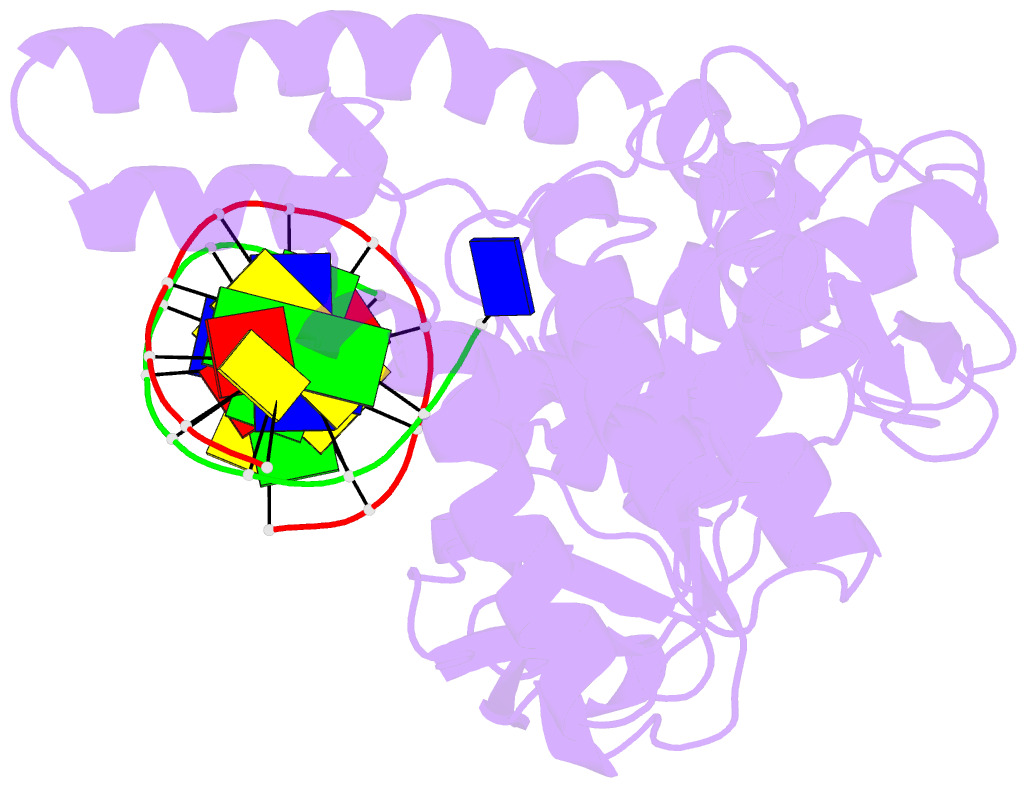
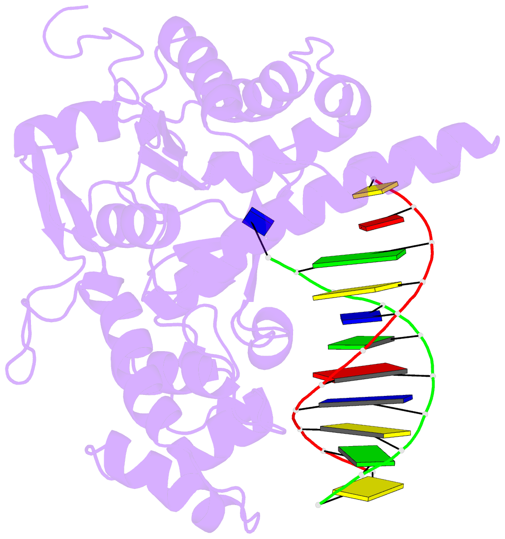
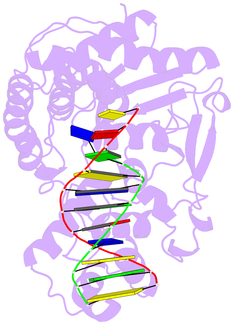
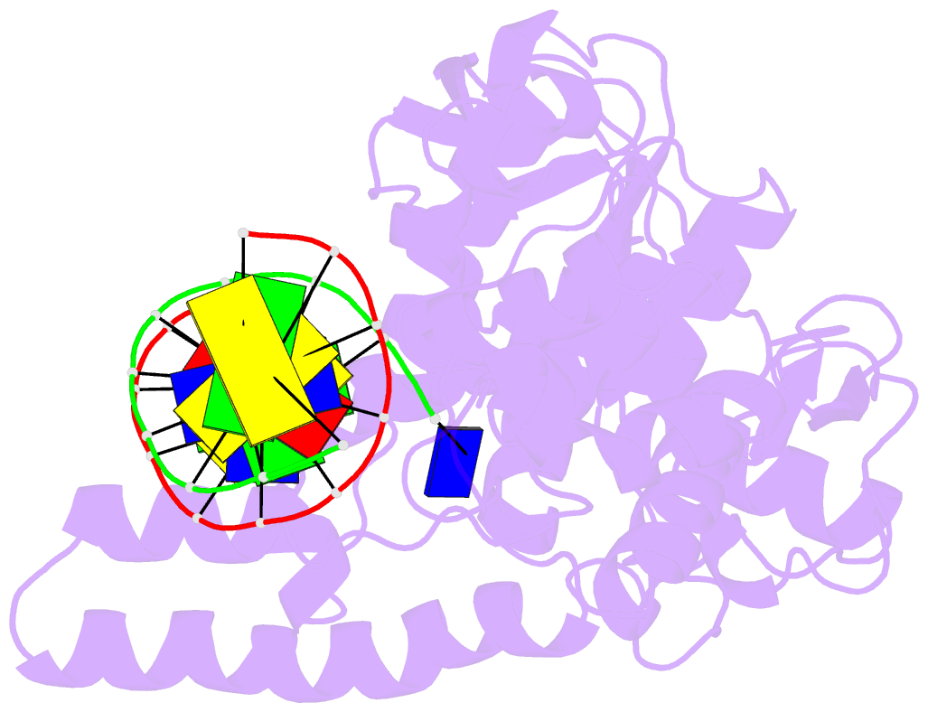
- Crystal structure of human exonuclease 1 exo1 (wt) in complex with DNA and mn2+ (complex iii)
- Reference
- Orans J, McSweeney EA, Iyer RR, Hast MA, Hellinga HW, Modrich P, Beese LS (2011): "Structures of human exonuclease 1 DNA complexes suggest a unified mechanism for nuclease family." Cell(Cambridge,Mass.), 145, 212-223. doi: 10.1016/j.cell.2011.03.005.
- Abstract
- Human exonuclease 1 (hExo1) plays important roles in DNA repair and recombination processes that maintain genomic integrity. It is a member of the 5' structure-specific nuclease family of exonucleases and endonucleases that includes FEN-1, XPG, and GEN1. We present structures of hExo1 in complex with a DNA substrate, followed by mutagenesis studies, and propose a common mechanism by which this nuclease family recognizes and processes diverse DNA structures. hExo1 induces a sharp bend in the DNA at nicks or gaps. Frayed 5' ends of nicked duplexes resemble flap junctions, unifying the mechanisms of endo- and exonucleolytic processing. Conformational control of a mobile region in the catalytic site suggests a mechanism for allosteric regulation by binding to protein partners. The relative arrangement of substrate binding sites in these enzymes provides an elegant solution to a complex geometrical puzzle of substrate recognition and processing.