Summary information and primary citation
- PDB-id
- 3qyx; SNAP-derived features in text and JSON formats;
DNAproDB
- Class
- transcription-DNA
- Method
- X-ray (3.75 Å)
- Summary
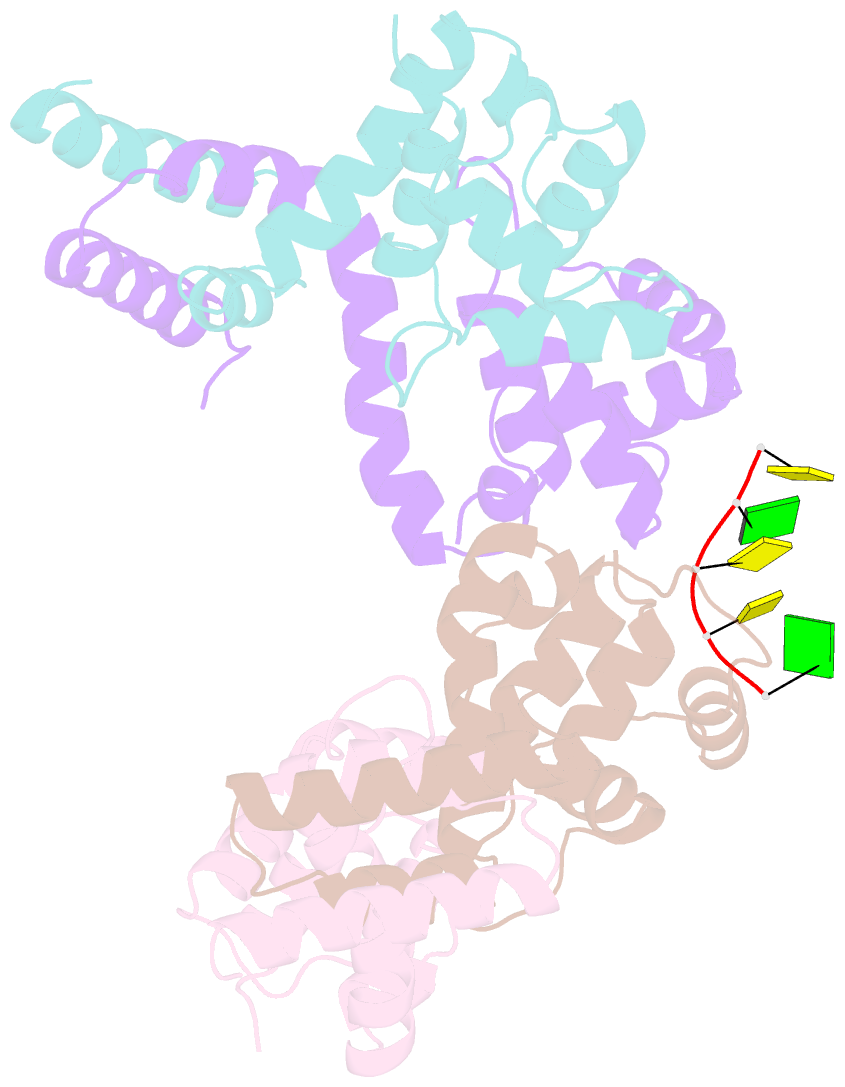
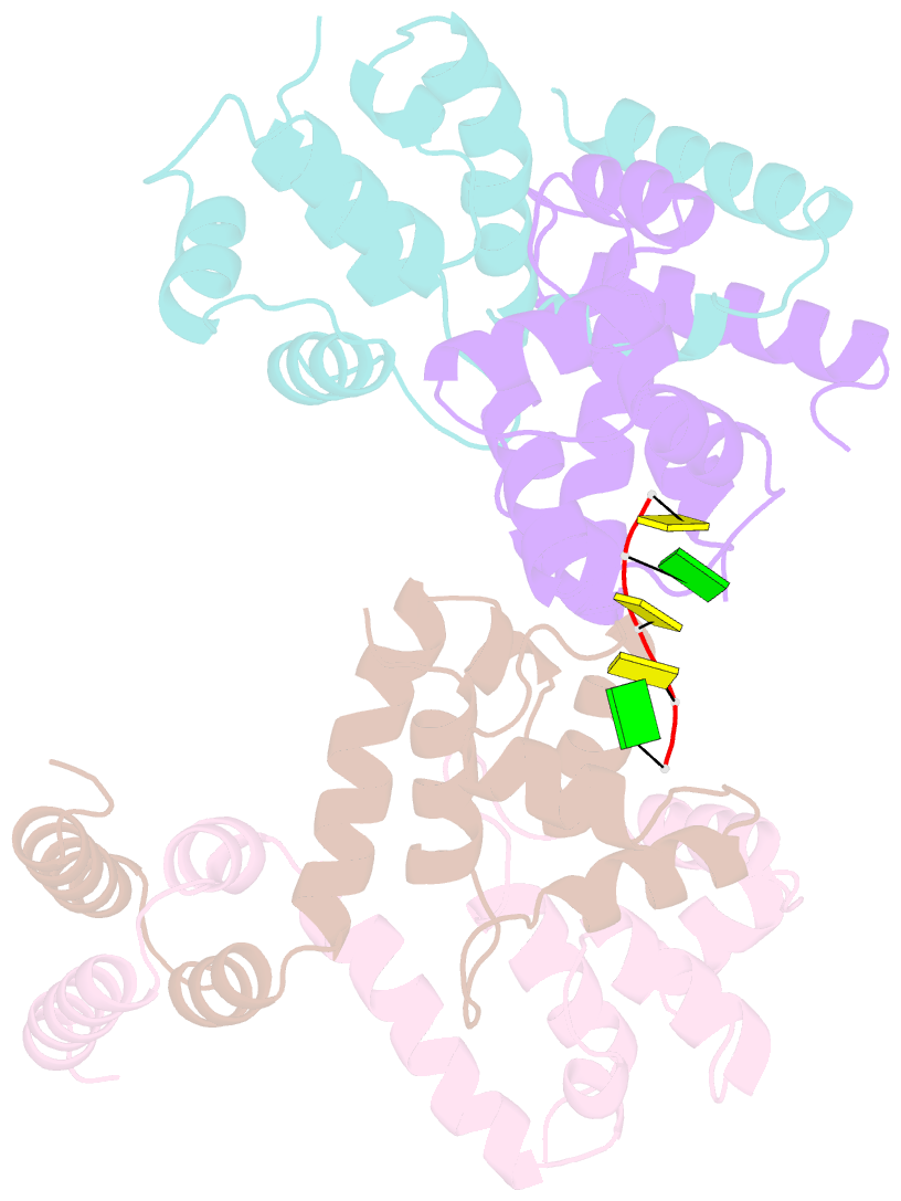
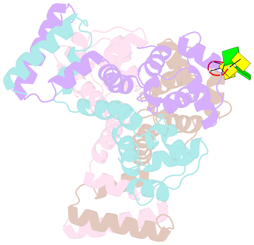
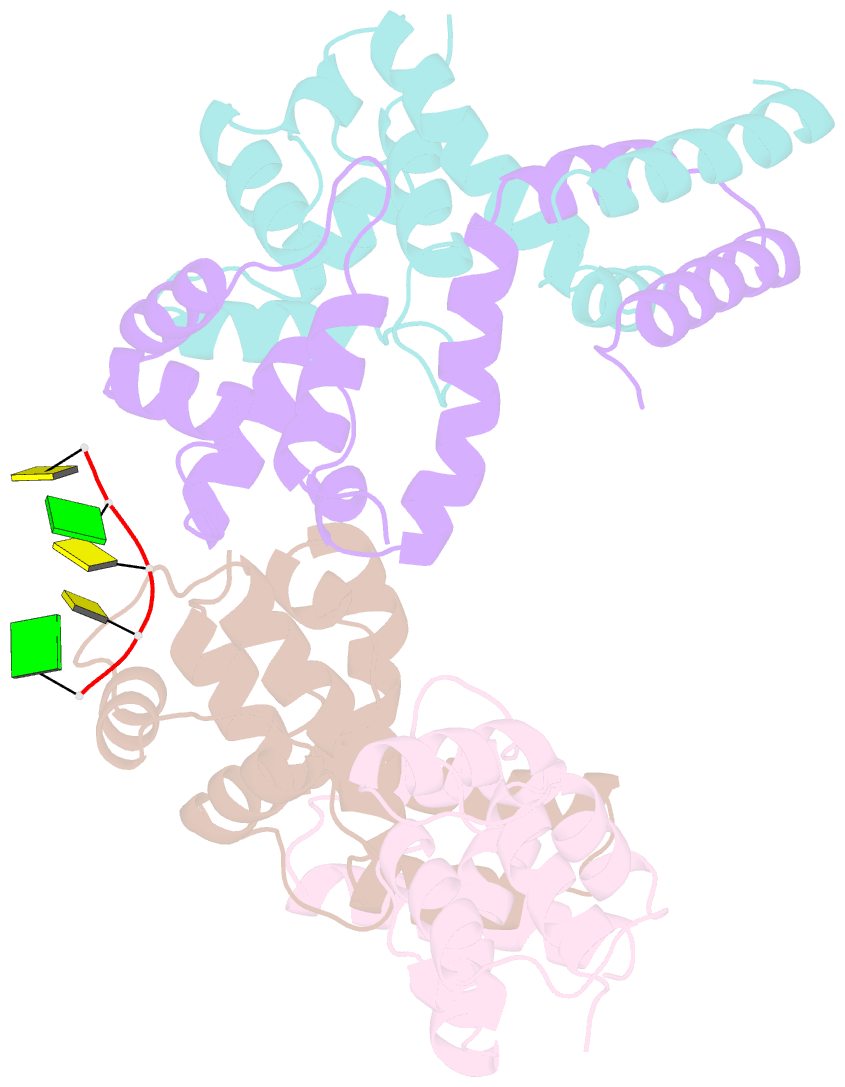
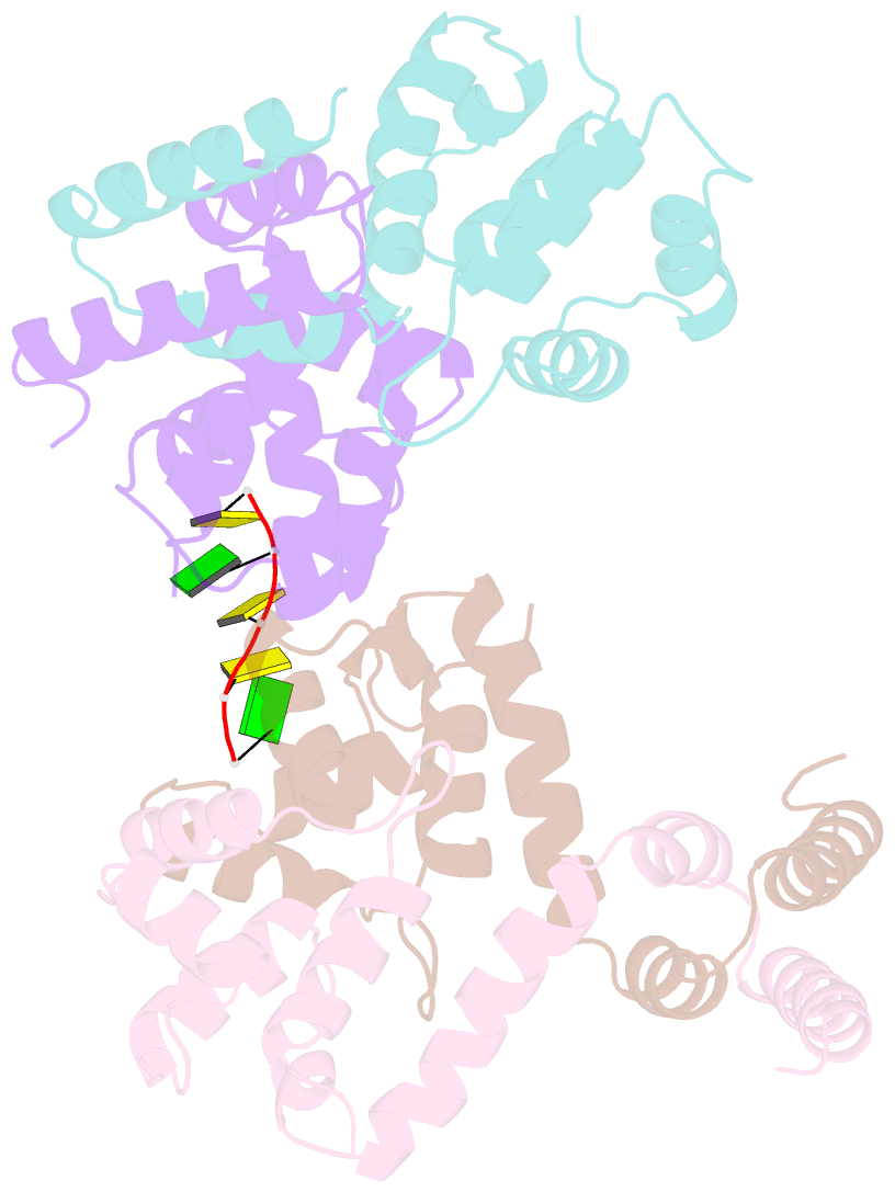
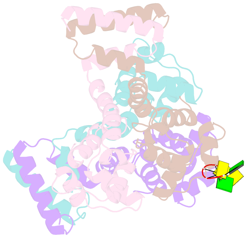
- Crystal structure of mycobacterium tuberculosis espr in complex with a small DNA fragment
- Reference
- Blasco B, Stenta M, Alonso-Sarduy L, Dietler G, Peraro MD, Cole ST, Pojer F (2011): "Atypical DNA recognition mechanism used by the EspR virulence regulator of Mycobacterium tuberculosis." Mol.Microbiol., 82, 251-264. doi: 10.1111/j.1365-2958.2011.07813.x.
- Abstract
- The human pathogen Mycobacterium tuberculosis requires the ESX-1 secretion system for full virulence. EspR plays a key role in ESX-1 regulation via direct binding and transcriptional activation of the espACD operon. Here, we describe the crystal structures of EspR, a C-terminally truncated form, EspRΔ10, as well as an EspR-DNA complex. EspR forms a dimer with each monomer containing an N-terminal helix-turn-helix DNA binding motif and an atypical C-terminal dimerization domain. Structural studies combined with footprinting experiments, atomic force microscopy and molecular dynamic simulations allow us to propose a model in which a dimer of EspR dimers is the minimal functional unit with two subunits binding two consecutive major grooves. The other two DNA binding domains are thus free to form higher-order oligomers and to bridge distant DNA sites in a cooperative way. These features are reminiscent of nucleoid-associated proteins and suggest a more general regulatory role for EspR than was previously suspected.