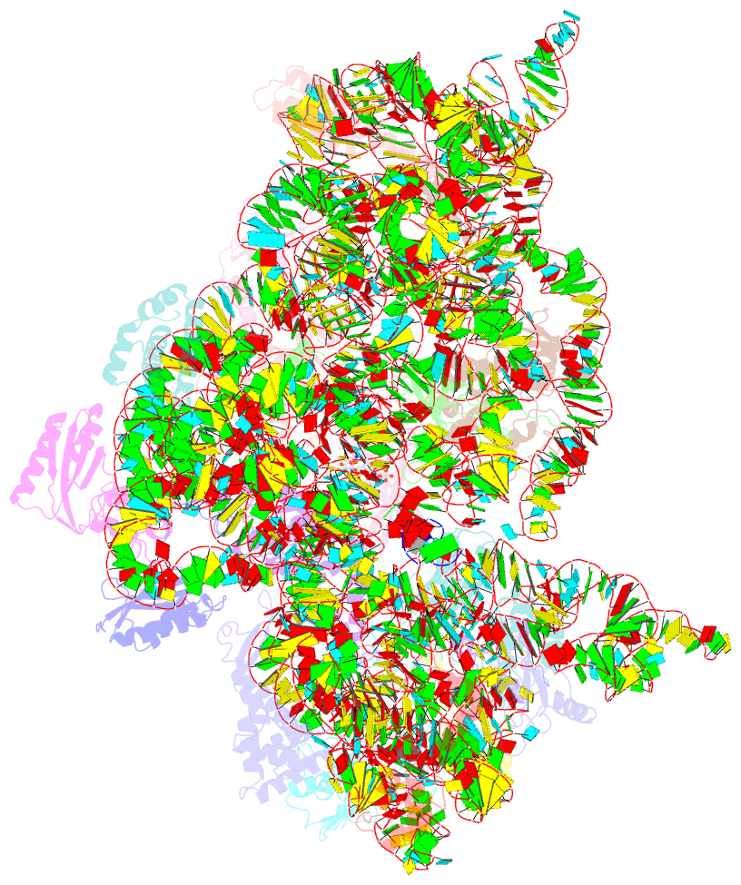
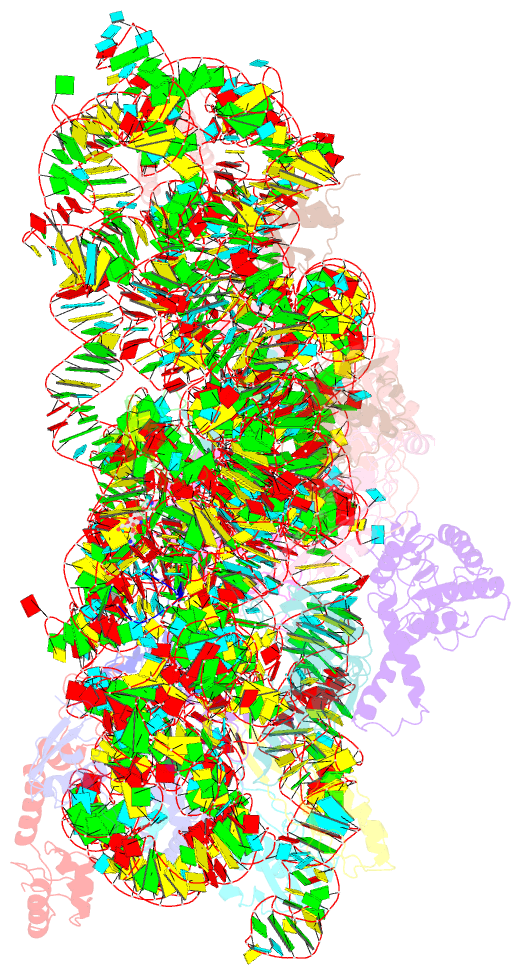
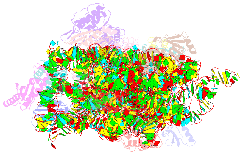
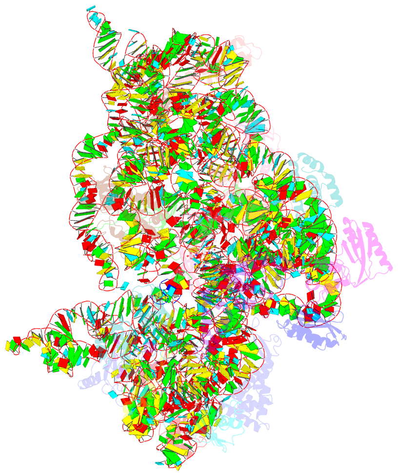
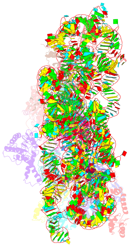
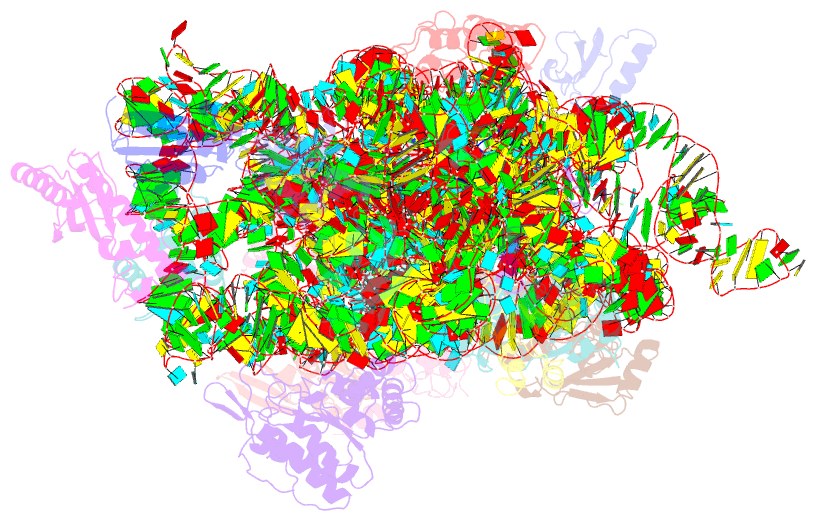
Summary information and primary citation
- PDB-id
- 3t1y; SNAP-derived features in text and JSON formats;
DNAproDB
- Class
- ribosome-antibiotic
- Method
- X-ray (2.8 Å)
- Summary
- Structure of the thermus thermophilus 30s ribosomal subunit complexed with a human anti-codon stem loop (hasl) of transfer RNA lysine 3 (trnalys3) bound to an mrna with an aag-codon in the a-site and paromomycin
- Reference
- Vendeix FA, Murphy FV, Cantara WA, Leszczynska G, Gustilo EM, Sproat B, Malkiewicz A, Agris PF (2012): "Human tRNA(Lys3)(UUU) Is Pre-Structured by Natural Modifications for Cognate and Wobble Codon Binding through Keto-Enol Tautomerism." J.Mol.Biol., 416, 467-485. doi: 10.1016/j.jmb.2011.12.048.
- Abstract
- Human tRNA(Lys3)(UUU) (htRNA(Lys3)(UUU)) decodes the lysine codons AAA and AAG during translation and also plays a crucial role as the primer for HIV-1 (human immunodeficiency virus type 1) reverse transcription. The posttranscriptional modifications 5-methoxycarbonylmethyl-2-thiouridine (mcm(5)s(2)U(34)), 2-methylthio-N(6)-threonylcarbamoyladenosine (ms(2)t(6)A(37)), and pseudouridine (Ψ(39)) in the tRNA's anticodon domain are critical for ribosomal binding and HIV-1 reverse transcription. To understand the importance of modified nucleoside contributions, we determined the structure and function of this tRNA's anticodon stem and loop (ASL) domain with these modifications at positions 34, 37, and 39, respectively (hASL(Lys3)(UUU)-mcm(5)s(2)U(34);ms(2)t(6)A(37);Ψ(39)). Ribosome binding assays in vitro revealed that the hASL(Lys3)(UUU)-mcm(5)s(2)U(34);ms(2)t(6)A(37);Ψ(39) bound AAA and AAG codons, whereas binding of the unmodified ASL(Lys3)(UUU) was barely detectable. The UV hyperchromicity, the circular dichroism, and the structural analyses indicated that Ψ(39) enhanced the thermodynamic stability of the ASL through base stacking while ms(2)t(6)A(37) restrained the anticodon to adopt an open loop conformation that is required for ribosomal binding. The NMR-restrained molecular-dynamics-derived solution structure revealed that the modifications provided an open, ordered loop for codon binding. The crystal structures of the hASL(Lys3)(UUU)-mcm(5)s(2)U(34);ms(2)t(6)A(37);Ψ(39) bound to the 30S ribosomal subunit with each codon in the A site showed that the modified nucleotides mcm(5)s(2)U(34) and ms(2)t(6)A(37) participate in the stability of the anticodon-codon interaction. Importantly, the mcm(5)s(2)U(34)·G(3) wobble base pair is in the Watson-Crick geometry, requiring unusual hydrogen bonding to G in which mcm(5)s(2)U(34) must shift from the keto to the enol form. The results unambiguously demonstrate that modifications pre-structure the anticodon as a key prerequisite for efficient and accurate recognition of cognate and wobble codons.