Summary information and primary citation
- PDB-id
- 3thw; SNAP-derived features in text and JSON formats;
DNAproDB
- Class
- DNA binding protein-DNA
- Method
- X-ray (3.09 Å)
- Summary
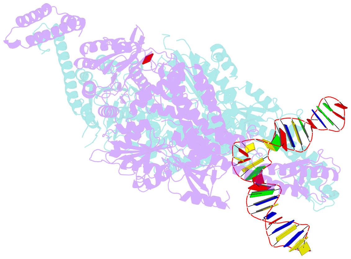
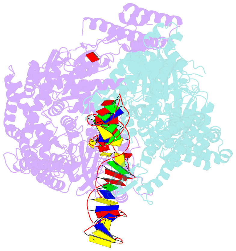
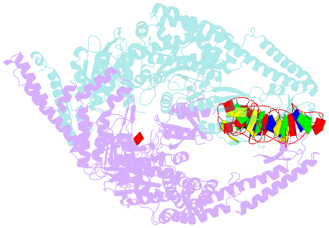
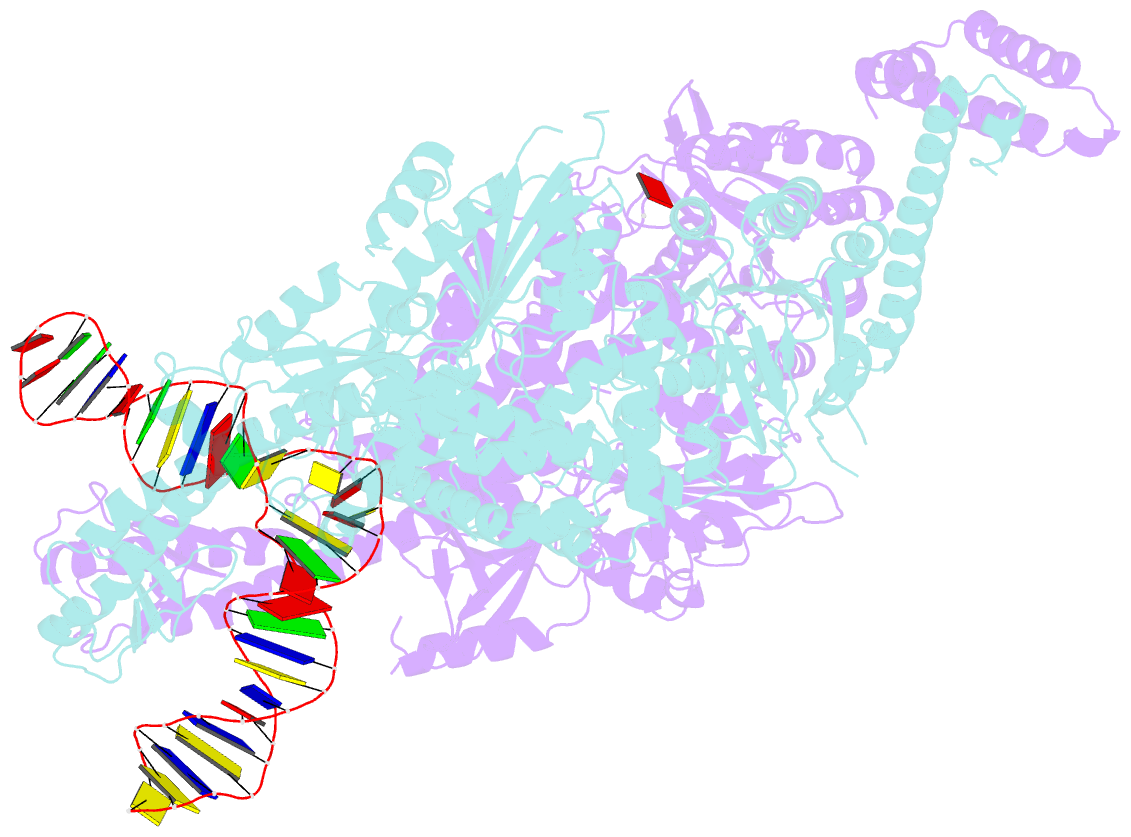
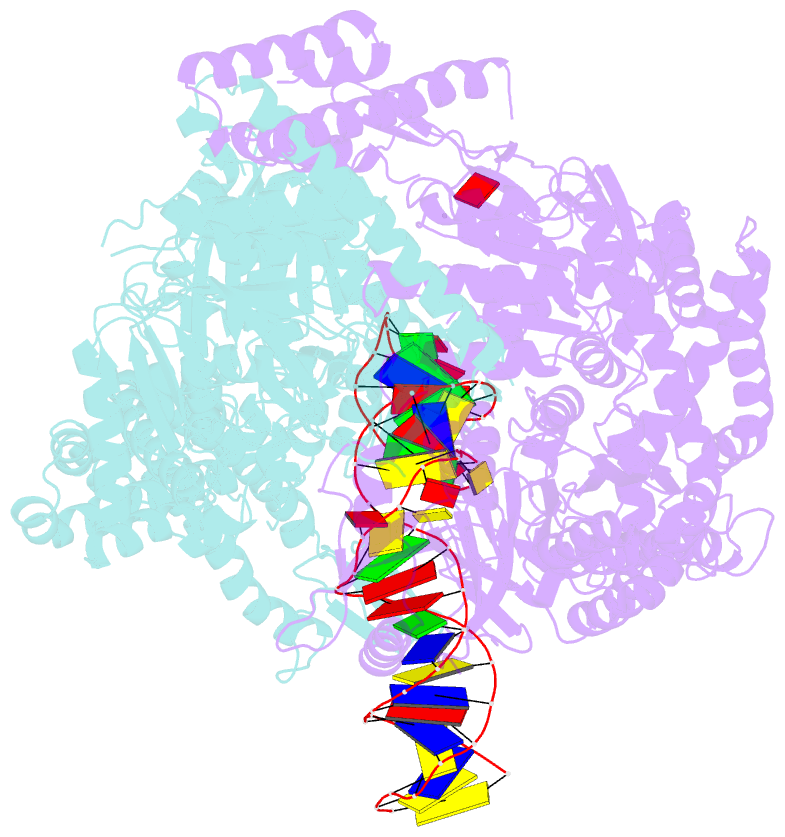
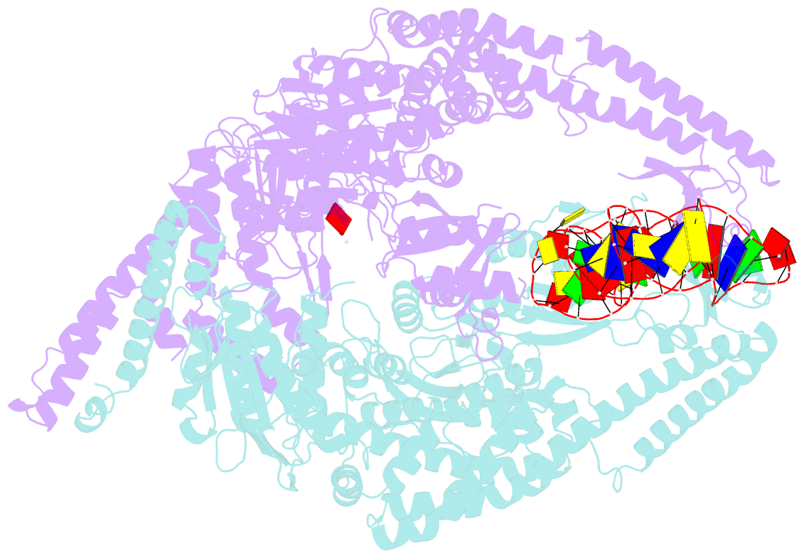
- Human mutsbeta complexed with an idl of 4 bases (loop4) and adp
- Reference
- Gupta S, Gellert M, Yang W (2012): "Mechanism of mismatch recognition revealed by human MutSbeta bound to unpaired DNA loops." Nat.Struct.Mol.Biol., 19, 72-78. doi: 10.1038/nsmb.2175.
- Abstract
- DNA mismatch repair corrects replication errors, thus reducing mutation rates and microsatellite instability. Genetic defects in this pathway cause Lynch syndrome and various cancers in humans. Binding of a mispaired or unpaired base by bacterial MutS and eukaryotic MutSα is well characterized. We report here crystal structures of human MutSβ in complex with DNA containing insertion-deletion loops (IDL) of two, three, four or six unpaired nucleotides. In contrast to eukaryotic MutSα and bacterial MutS, which bind the base of a mismatched nucleotide, MutSβ binds three phosphates in an IDL. DNA is severely bent at the IDL; unpaired bases are flipped out into the major groove and partially exposed to solvent. A normal downstream base pair can become unpaired; a single unpaired base can thereby be converted to an IDL of two nucleotides and recognized by MutSβ. The C-terminal dimerization domains form an integral part of the MutS structure and coordinate asymmetrical ATP hydrolysis by Msh2 and Msh3 with mismatch binding to signal for repair.