Summary information and primary citation
- PDB-id
- 3u3w; SNAP-derived features in text and JSON formats;
DNAproDB
- Class
- transcription activator-DNA
- Method
- X-ray (2.4 Å)
- Summary
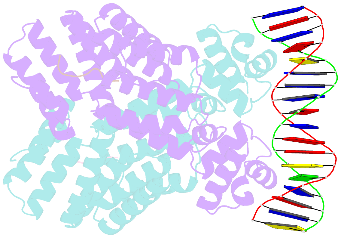
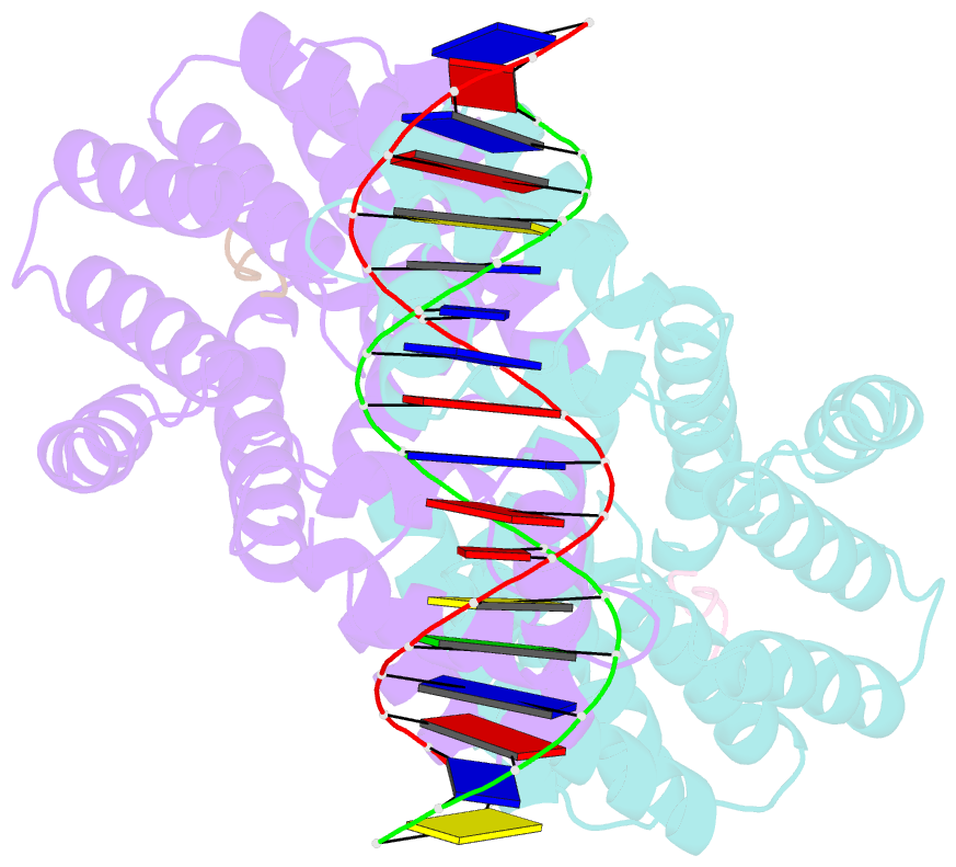
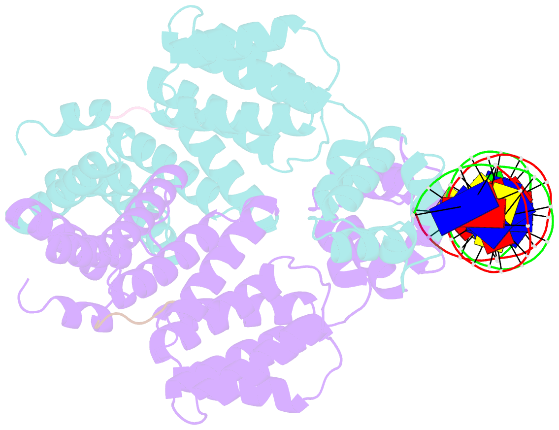
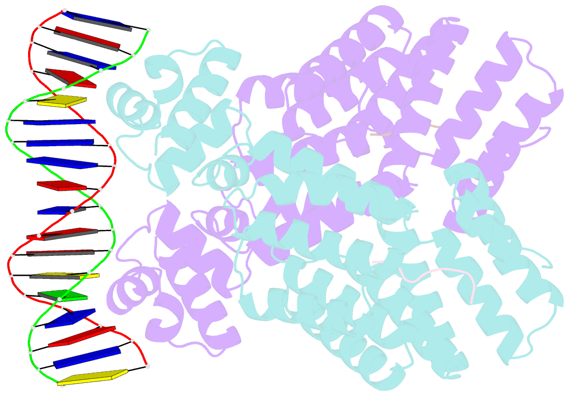
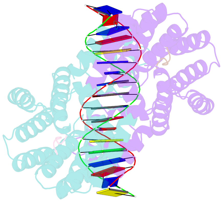
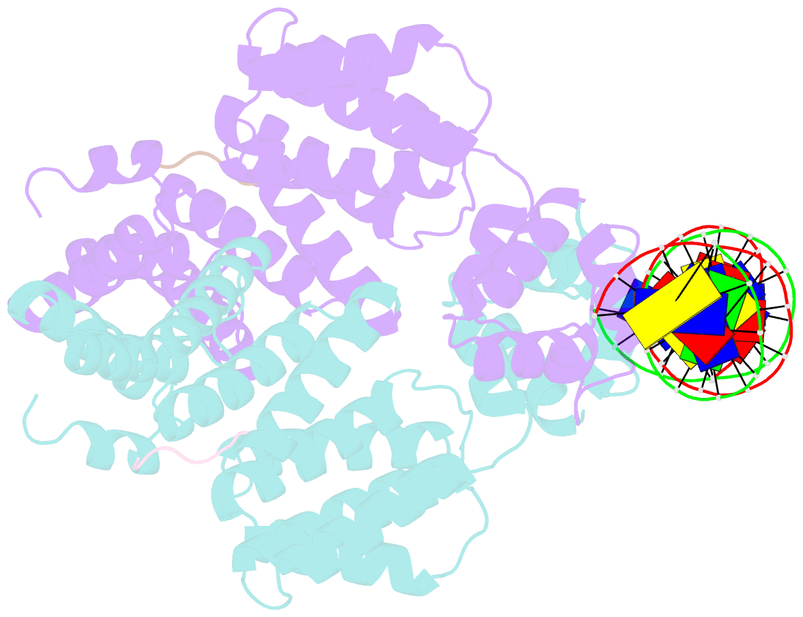
- Crystal structure of bacillus thuringiensis plcr in complex with the peptide papr7 and DNA
- Reference
- Grenha R, Slamti L, Nicaise M, Refes Y, Lereclus D, Nessler S (2013): "Structural basis for the activation mechanism of the PlcR virulence regulator by the quorum-sensing signal peptide PapR." Proc.Natl.Acad.Sci.USA, 110, 1047-1052. doi: 10.1073/pnas.1213770110.
- Abstract
- The quorum-sensing regulator PlcR is the master regulator of most known virulence factors in Bacillus cereus. It is a helix-turn-helix (HTH)-type transcription factor activated upon binding of its cognate signaling peptide PapR on a tetratricopeptide repeat-type regulatory domain. The structural and functional properties of PlcR have defined a new family of sensor regulators, called the RNPP family (for Rap, NprR, PrgX, and PlcR), in Gram-positive bacteria. To fully understand the activation mechanism of PlcR, we took a closer look at the conformation changes induced upon binding of PapR and of its target DNA, known as PlcR-box. For that purpose we have determined the structures of the apoform of PlcR (Apo PlcR) and of the ternary complex of PlcR with PapR and the PlcR-box from the plcA promoter. Comparison of the apoform of PlcR with the previously published structure of the PlcR-PapR binary complex shows how a small conformational change induced in the C-terminal region of the tetratricopeptide repeat (TPR) domain upon peptide binding propagates via the linker helix to the N-terminal HTH DNA-binding domain. Further comparison with the PlcR-PapR-DNA ternary complex shows how the activation of the PlcR dimer allows the linker helix to undergo a drastic conformational change and subsequent proper positioning of the HTH domains in the major groove of the two half sites of the pseudopalindromic PlcR-box. Together with random mutagenesis experiments and interaction measurements using peptides from distinct pherogroups, this structural analysis allows us to propose a molecular mechanism for this functional switch.