Summary information and primary citation
- PDB-id
- 3ugm; SNAP-derived features in text and JSON formats;
DNAproDB
- Class
- transcription-DNA
- Method
- X-ray (3.0 Å)
- Summary
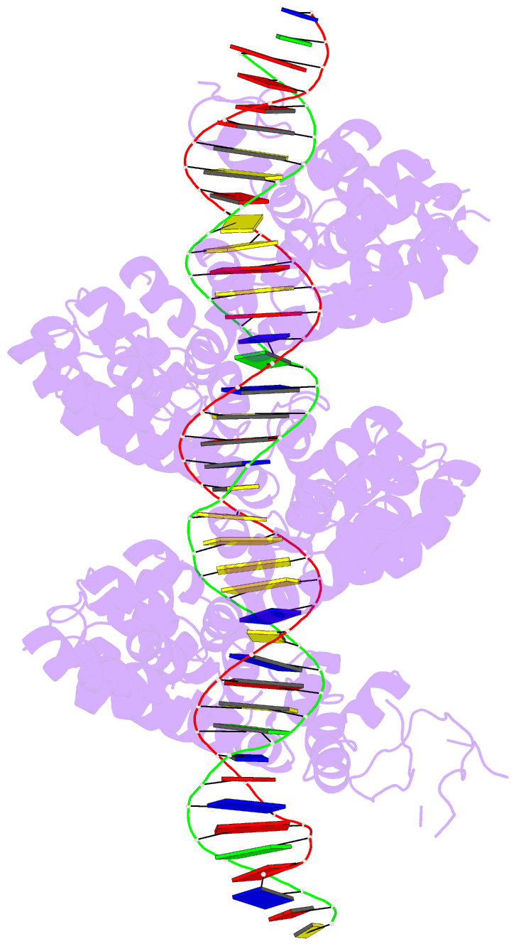
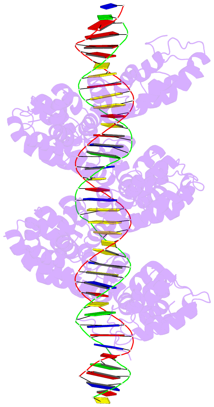
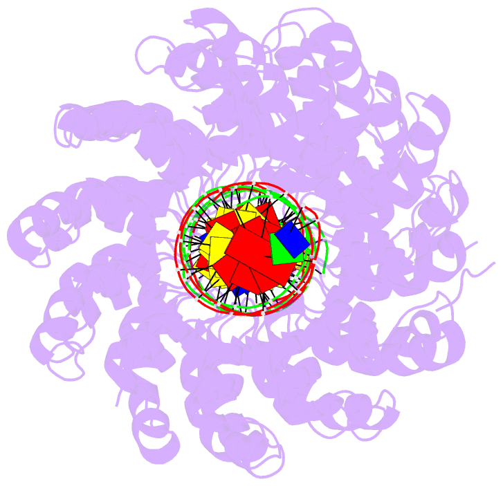
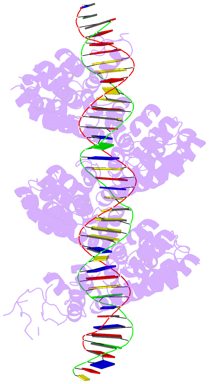
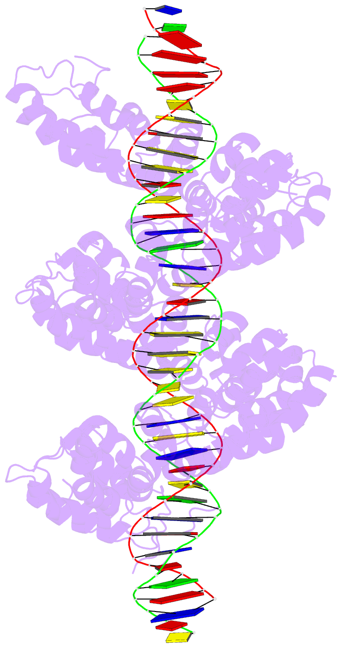
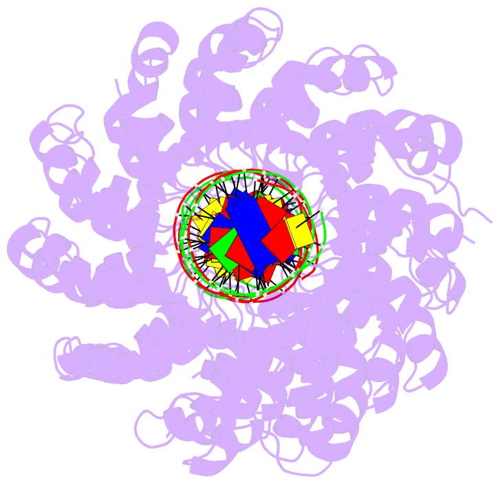
- Structure of tal effector pthxo1 bound to its DNA target
- Reference
- Mak AN, Bradley P, Cernadas RA, Bogdanove AJ, Stoddard BL (2012): "The Crystal Structure of TAL Effector PthXo1 Bound to Its DNA Target." Science, 335, 716-719. doi: 10.1126/science.1216211.
- Abstract
- DNA recognition by TAL effectors is mediated by tandem repeats, each 33 to 35 residues in length, that specify nucleotides via unique repeat-variable diresidues (RVDs). The crystal structure of PthXo1 bound to its DNA target was determined by high-throughput computational structure prediction and validated by heavy-atom derivatization. Each repeat forms a left-handed, two-helix bundle that presents an RVD-containing loop to the DNA. The repeats self-associate to form a right-handed superhelix wrapped around the DNA major groove. The first RVD residue forms a stabilizing contact with the protein backbone, while the second makes a base-specific contact to the DNA sense strand. Two degenerate amino-terminal repeats also interact with the DNA. Containing several RVDs and noncanonical associations, the structure illustrates the basis of TAL effector-DNA recognition.