Summary information and primary citation
- PDB-id
- 3w2a; SNAP-derived features in text and JSON formats;
DNAproDB
- Class
- transcription-DNA
- Method
- X-ray (2.775 Å)
- Summary
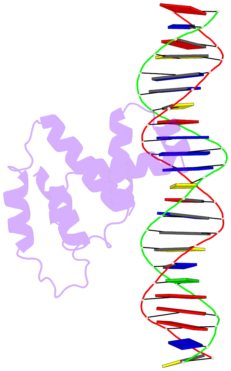
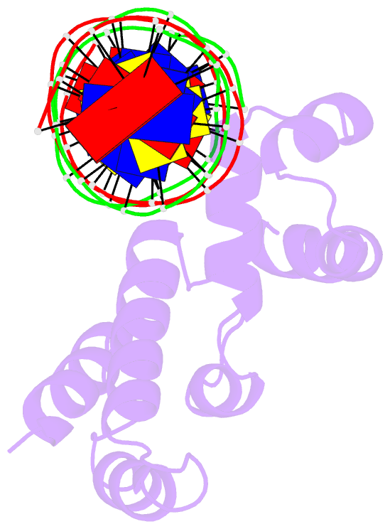
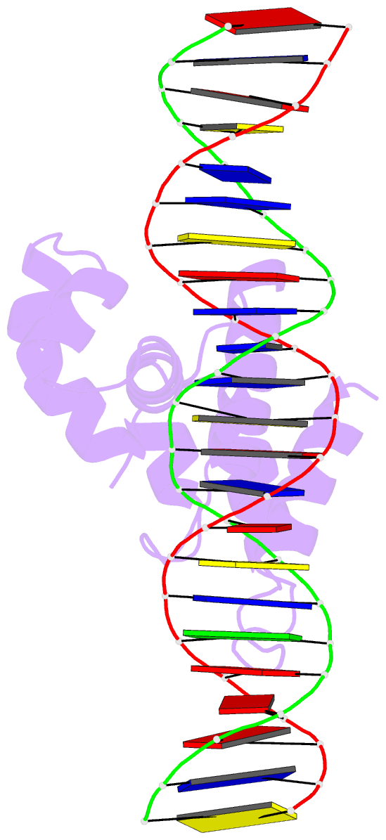
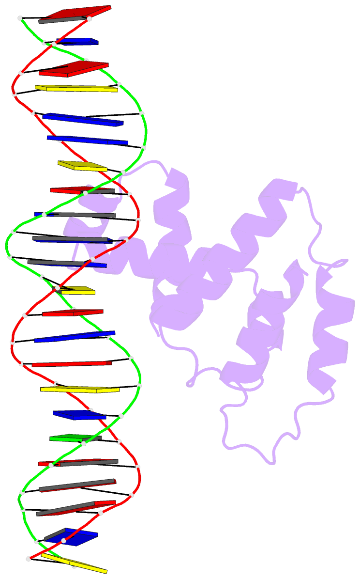
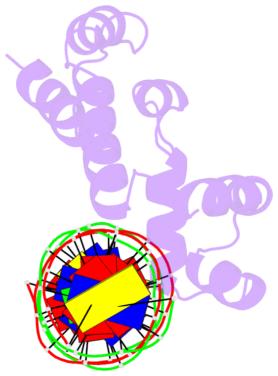
- Crystal structure of virb core domain complexed with the cis-acting site upstream icsp promoter
- Reference
- Gao XP, Zou TT, Mu ZX, Qin B, Yang J, Waltersperger S, Wang MT, Cui S, Jin Q (2013): "Structural insights into VirB-DNA complexes reveal mechanism of transcriptional activation of virulence genes." Nucleic Acids Res., 41, 10529-10541. doi: 10.1093/nar/gkt748.
- Abstract
- VirB activates transcription of virulence genes in Shigella flexneri by alleviating heat-stable nucleoid-structuring protein-mediated promoter repression. VirB is unrelated to the conventional transcriptional regulators, but homologous to the plasmid partitioning proteins. We determined the crystal structures of VirB HTH domain bound by the cis-acting site containing the inverted repeat, revealing that the VirB-DNA complex is related to ParB-ParS-like complexes, presenting an example that a ParB-like protein acts exclusively in transcriptional regulation. The HTH domain of VirB docks DNA major groove and provides multiple contacts to backbone and bases, in which the only specific base readout is mediated by R167. VirB only recognizes one half site of the inverted repeats containing the most matches to the consensus for VirB binding. The binding of VirB induces DNA conformational changes and introduces a bend at an invariant A-tract segment in the cis-acting site, suggesting a role of DNA remodeling. VirB exhibits positive cooperativity in DNA binding that is contributed by the C-terminal domain facilitating VirB oligomerization. The isolated HTH domain only confers partial DNA specificity. Additional determinants for sequence specificity may reside in N- or C-terminal domains. Collectively, our findings support and extend a previously proposed model for relieving heat-stable nucleoid-structuring protein-mediated repression by VirB.