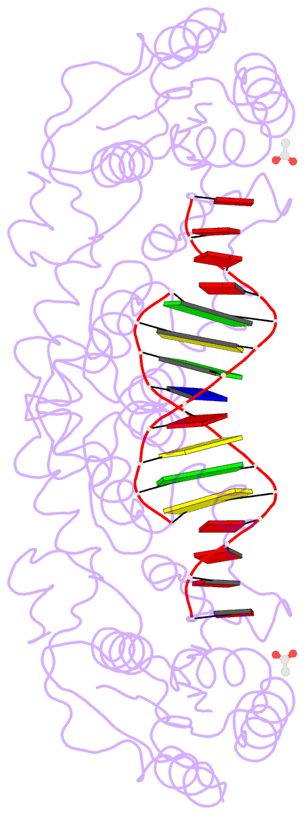
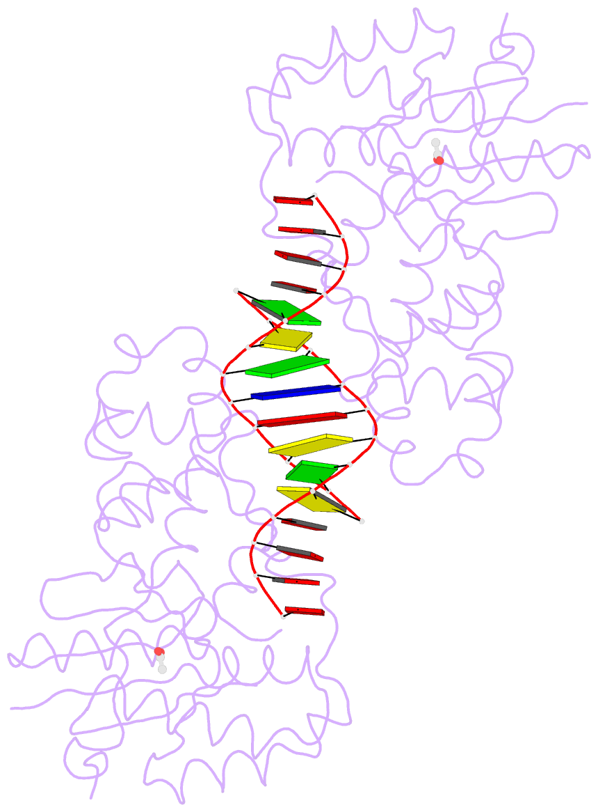
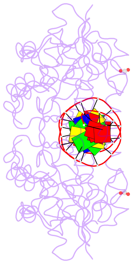
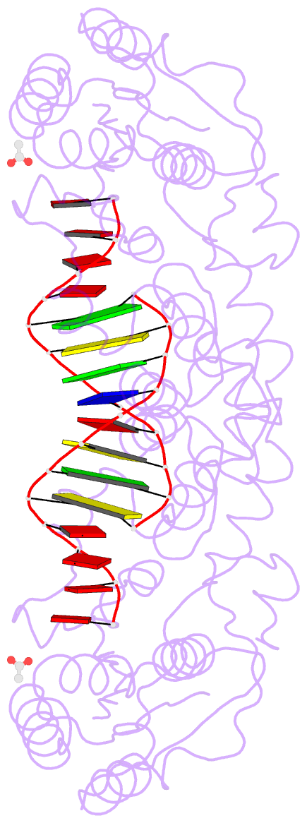
Summary information and primary citation
- PDB-id
- 3zdb; SNAP-derived features in text and JSON formats;
DNAproDB
- Class
- hydrolase-DNA
- Method
- X-ray (1.47 Å)
- Summary
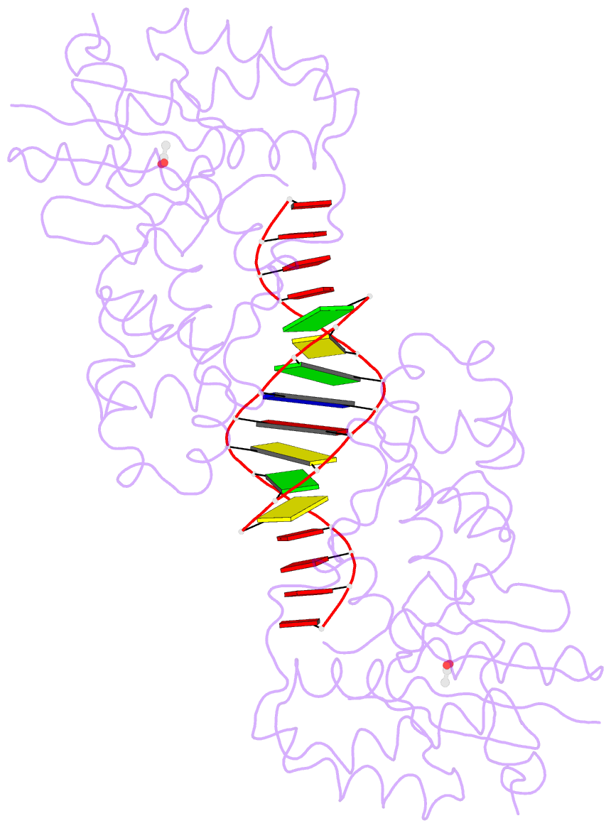
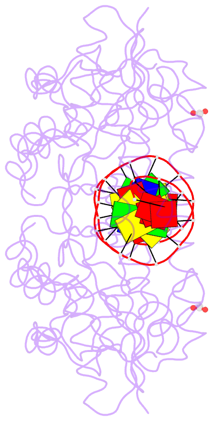
- Structure of e. coli exoix in complex with the palindromic 5ov4 DNA oligonucleotide, di-magnesium and potassium
- Reference
- Anstey-Gilbert CS, Hemsworth GR, Flemming CS, Hodskinson MRG, Zhang J, Sedelnikova SE, Stillman TJ, Sayers JR, Artymiuk PJ (2013): "The Structure of E. Coli Exoix - Implications for DNA Binding and Catalysis in Flap Endonucleases." Nucleic Acids Res., 41, 8357. doi: 10.1093/NAR/GKT591.
- Abstract
- Escherichia coli Exonuclease IX (ExoIX), encoded by the xni gene, was the first identified member of a novel subfamily of ubiquitous flap endonucleases (FENs), which possess only one of the two catalytic metal-binding sites characteristic of other FENs. We have solved the first structure of one of these enzymes, that of ExoIX itself, at high resolution in DNA-bound and DNA-free forms. In the enzyme-DNA cocrystal, the single catalytic site binds two magnesium ions. The structures also reveal a binding site in the C-terminal domain where a potassium ion is directly coordinated by five main chain carbonyl groups, and we show this site is essential for DNA binding. This site resembles structurally and functionally the potassium sites in the human FEN1 and exonuclease 1 enzymes. Fluorescence anisotropy measurements and the crystal structures of the ExoIX:DNA complexes show that this potassium ion interacts directly with a phosphate diester in the substrate DNA.