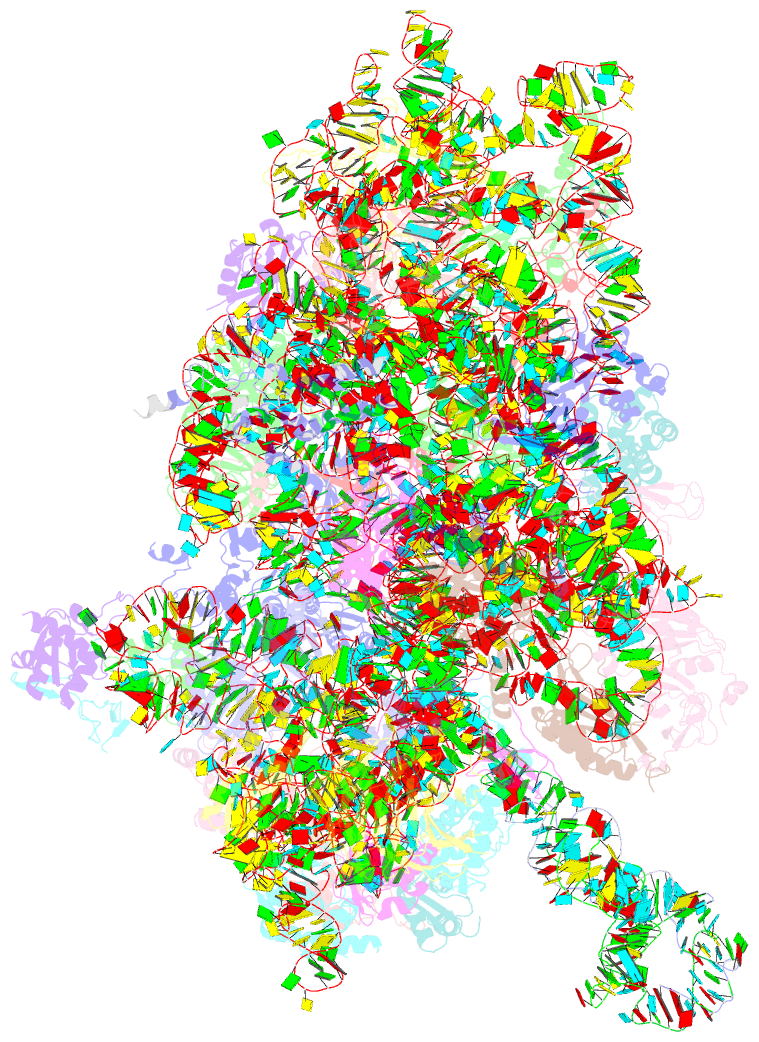
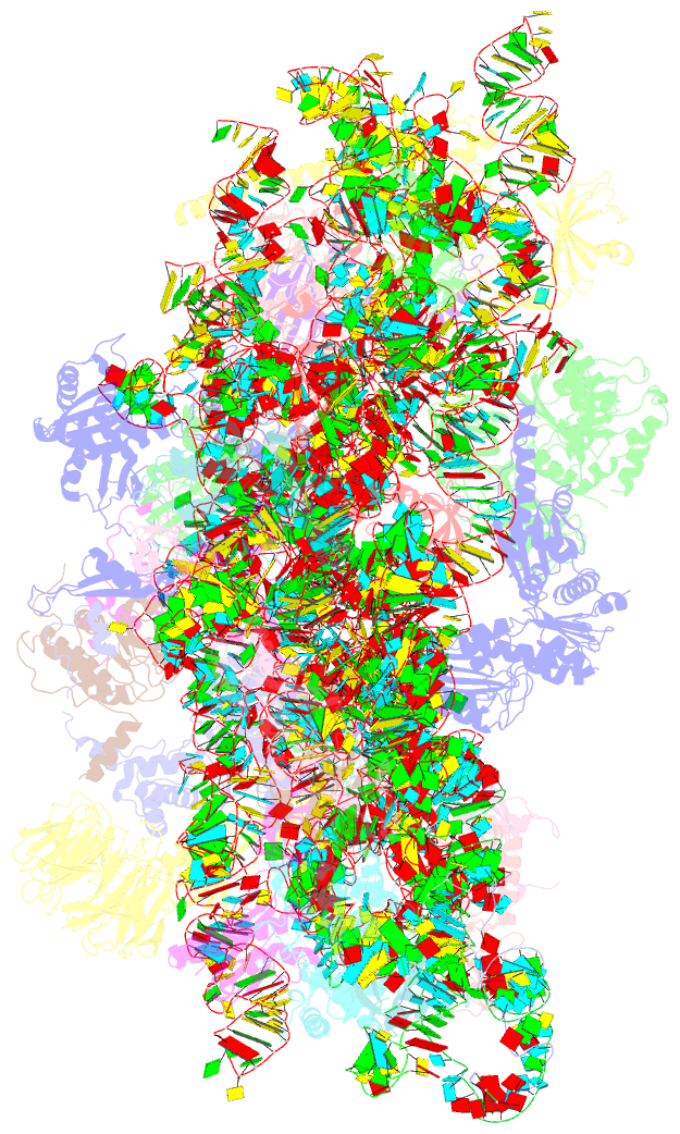
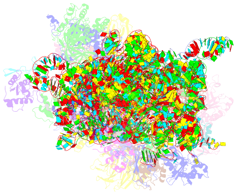
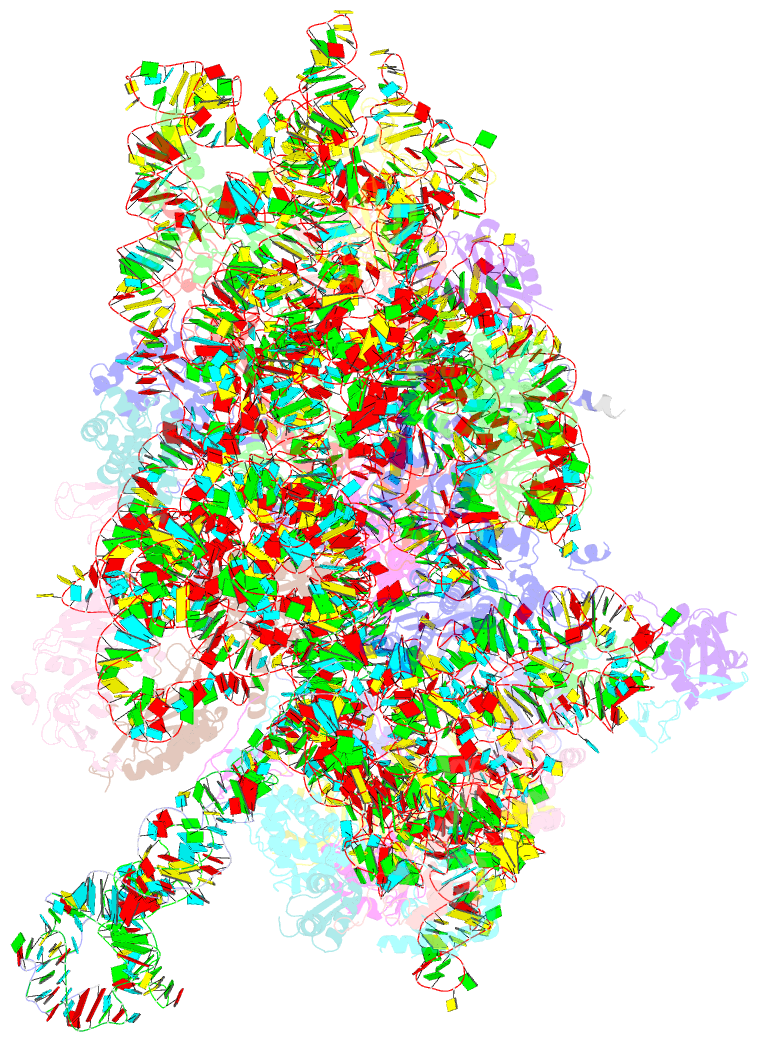
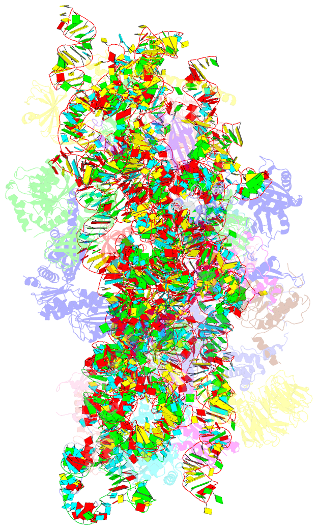
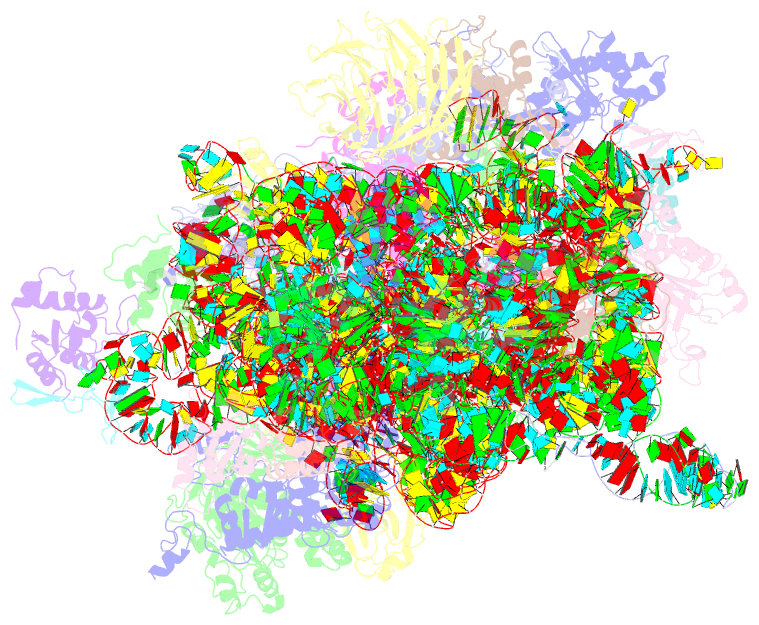
Summary information and primary citation
- PDB-id
- 4d61; SNAP-derived features in text and JSON formats;
DNAproDB
- Class
- ribosome
- Method
- cryo-EM (9.0 Å)
- Summary
- cryo-EM structures of ribosomal 80s complexes with termination factors and cricket paralysis virus ires reveal the ires in the translocated state
- Reference
- Muhs M, Hilal T, Mielke T, Skabkin MA, Sanbonmatsu KY, Pestova TV, Spahn CMT (2015): "Cryo-Em of Ribosomal 80S Complexes with Termination Factors Reveals the Translocated Cricket Paralysis Virus Ires." Mol.Cell, 57, 422. doi: 10.1016/J.MOLCEL.2014.12.016.
- Abstract
- The cricket paralysis virus (CrPV) uses an internal ribosomal entry site (IRES) to hijack the ribosome. In a remarkable RNA-based mechanism involving neither initiation factor nor initiator tRNA, the CrPV IRES jumpstarts translation in the elongation phase from the ribosomal A site. Here, we present cryoelectron microscopy (cryo-EM) maps of 80S⋅CrPV-STOP ⋅ eRF1 ⋅ eRF3 ⋅ GMPPNP and 80S⋅CrPV-STOP ⋅ eRF1 complexes, revealing a previously unseen binding state of the IRES and directly rationalizing that an eEF2-dependent translocation of the IRES is required to allow the first A-site occupation. During this unusual translocation event, the IRES undergoes a pronounced conformational change to a more stretched conformation. At the same time, our structural analysis provides information about the binding modes of eRF1 ⋅ eRF3 ⋅ GMPPNP and eRF1 in a minimal system. It shows that neither eRF3 nor ABCE1 are required for the active conformation of eRF1 at the intersection between eukaryotic termination and recycling.