Summary information and primary citation
- PDB-id
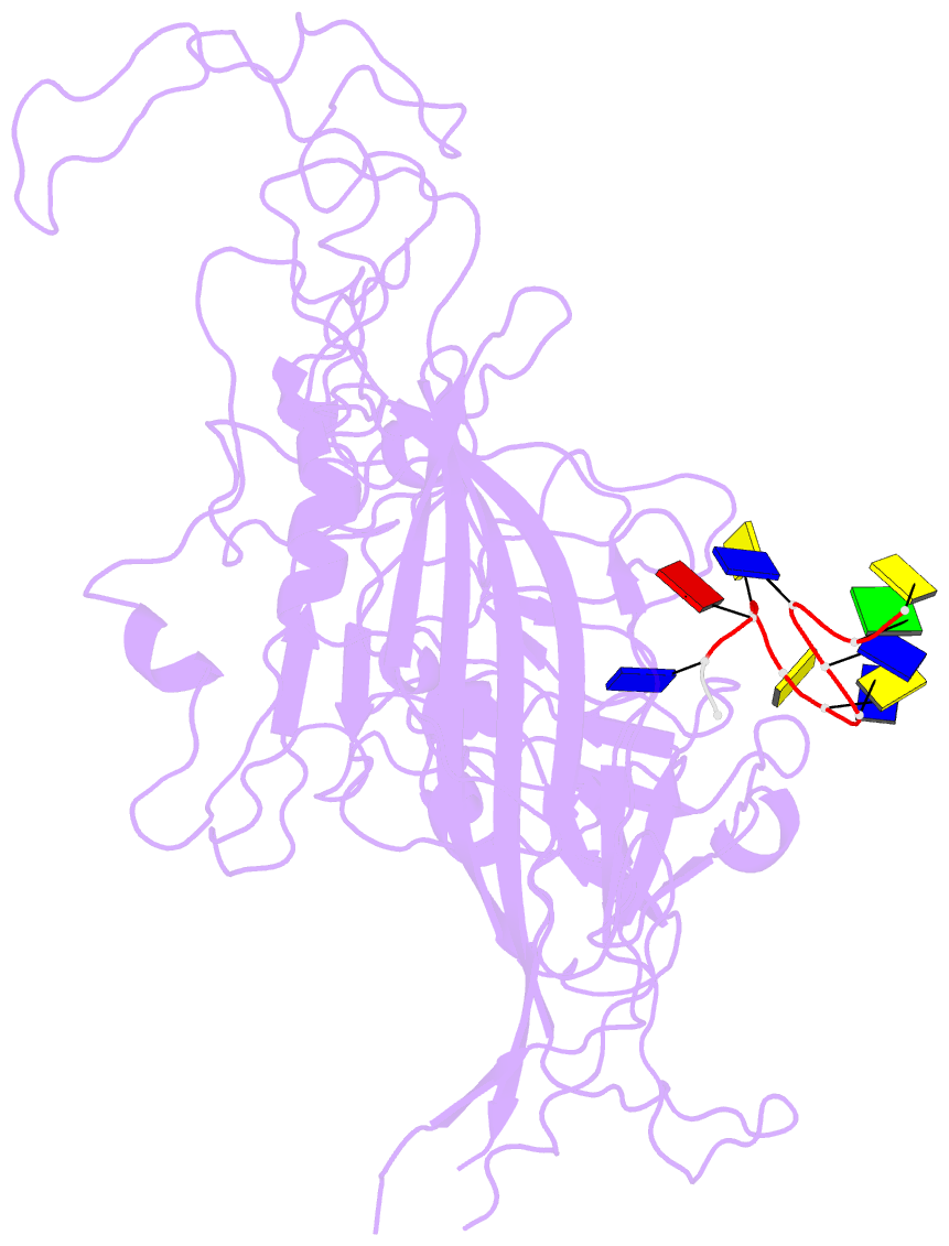
- 4dpv; SNAP-derived features in text and JSON formats;
DNAproDB
- Class
- virus-DNA
- Method
- X-ray (2.9 Å)
- Summary
- Parvovirus-DNA complex
- Reference
- Xie Q, Chapman MS (1996): "Canine parvovirus capsid structure, analyzed at 2.9 A resolution." J.Mol.Biol., 264, 497-520. doi: 10.1006/jmbi.1996.0657.
- Abstract
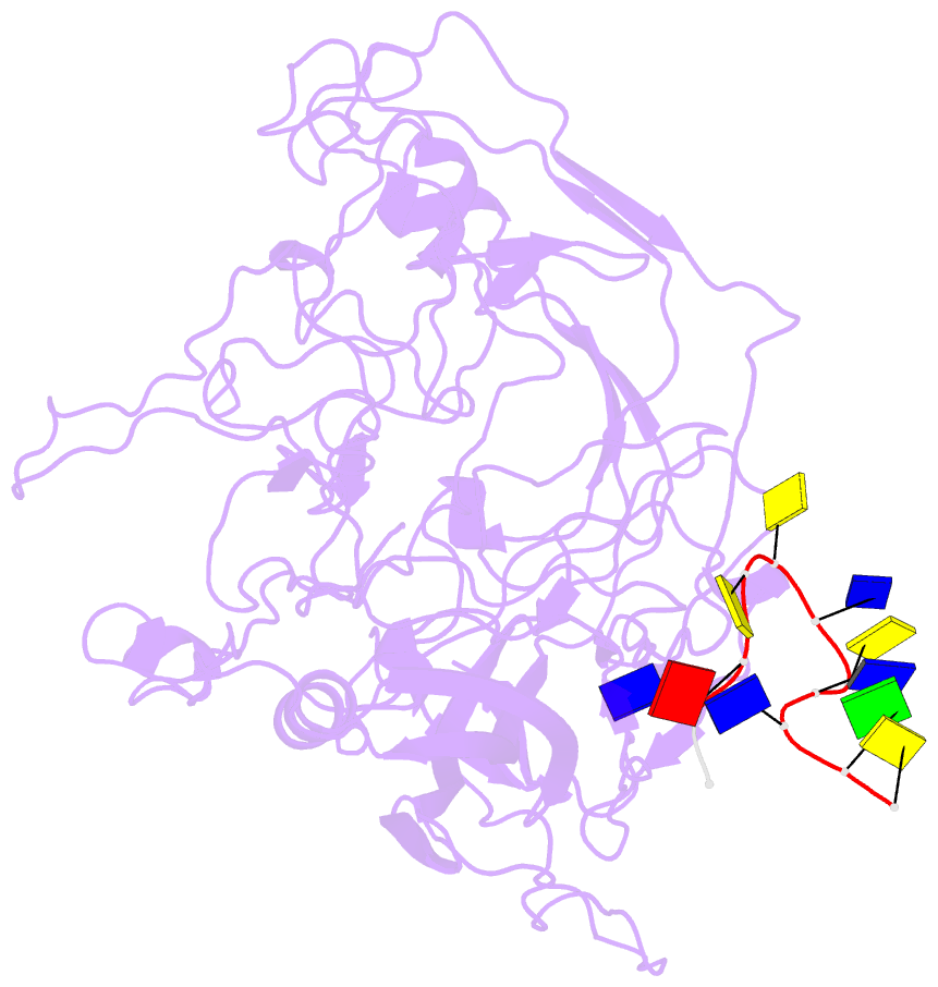
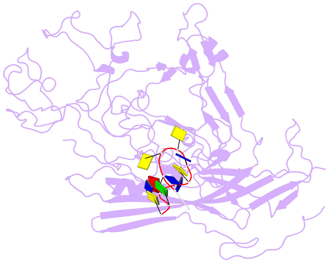
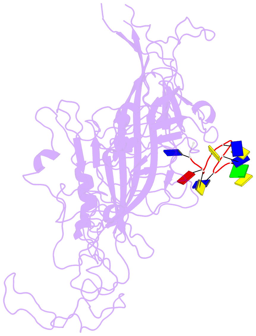
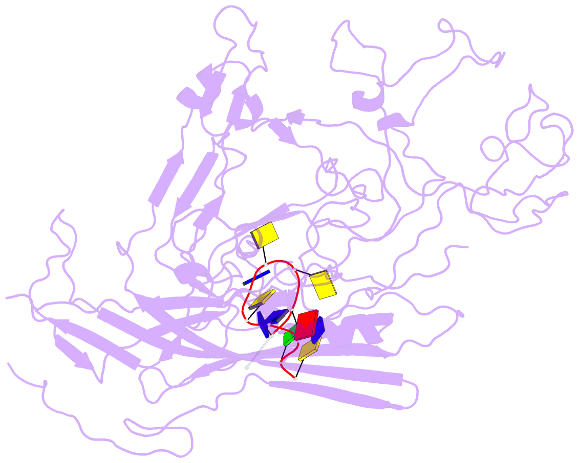
- The DNA-containing capsid of canine parvovirus (CPV) is analyzed following atomic refinement at 2.9 A resolution. The capsid contains 60 copies of the capsid protein related by icosahedral symmetry. The atomic model has been extended from the first residue (Gly37) of the unrefined 3.25 A structure towards the N terminus. The electron density shows that approximately 87% of the capsid proteins have N termini on the inside of the capsid, but for approximately 13%, the polypeptide starts on the outside and runs through one of the pores surrounding each 5-fold axis, explaining apparently conflicting antigenic data. Analysis of potential hydrogen bonds reveals approximately 50% more secondary structure than previously apparent. Most of the additional secondary structure are in the 71 and 221 residue-long loop insertions between beta-strands E and F and G and H, forming subunit-bridging sheets that likely add specificity to assembly interactions. Structural analysis of the extensive subunit interactions around the 3-fold axes shows that assembly is a multistep process with loops intertwining following initial contact. Estimated free energies of association suggest that the formation of 3 and 5-fold contacts likely takes precedence over 2-fold interactions. Energies for initial association into trimers or pentamers would be similar, but the intertwining of loops about the 3-fold axis adds an additional large activation barrier to dissociation. Analysis of the surfaces of the assembled capsid shows a surprising lack of basic amino acids that might have been expected to interact with the negatively charged phosphoribose backbone of the DNA. Instead, uncharged polar and van der Waal's interactions predominate in the packaging of single-stranded DNA into the capsid.