Summary information and primary citation
- PDB-id
- 4fpv; SNAP-derived features in text and JSON formats;
DNAproDB
- Class
- hydrolase-DNA
- Method
- X-ray (1.73 Å)
- Summary
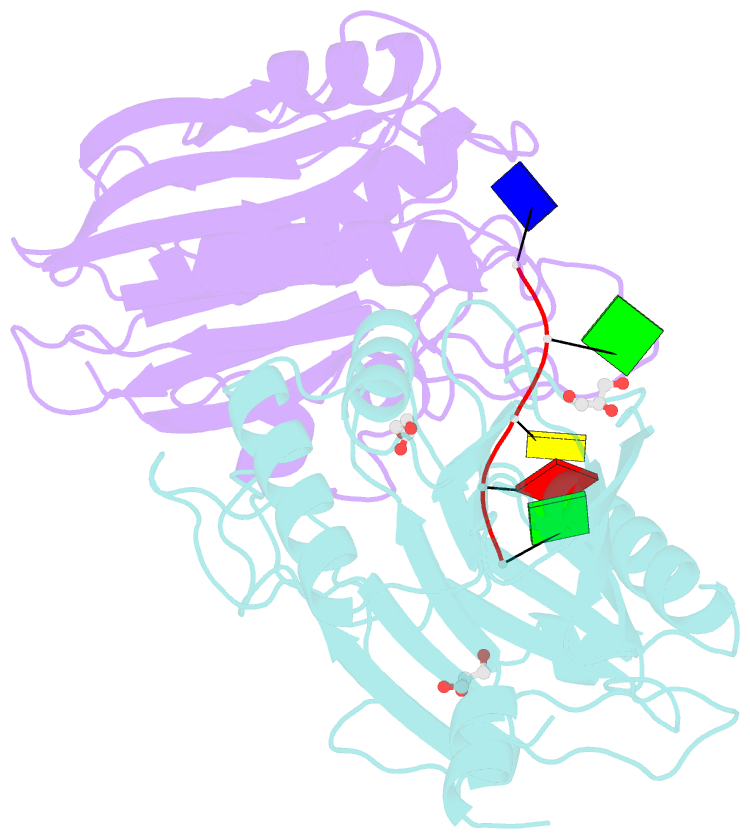
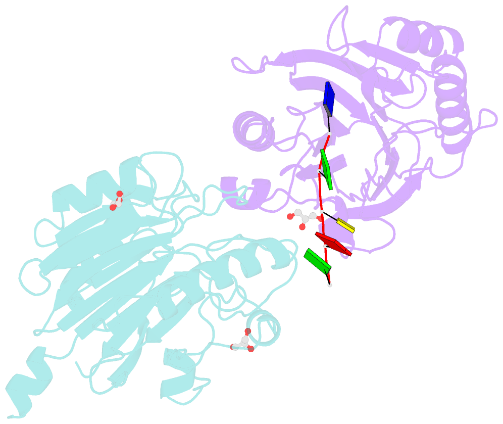
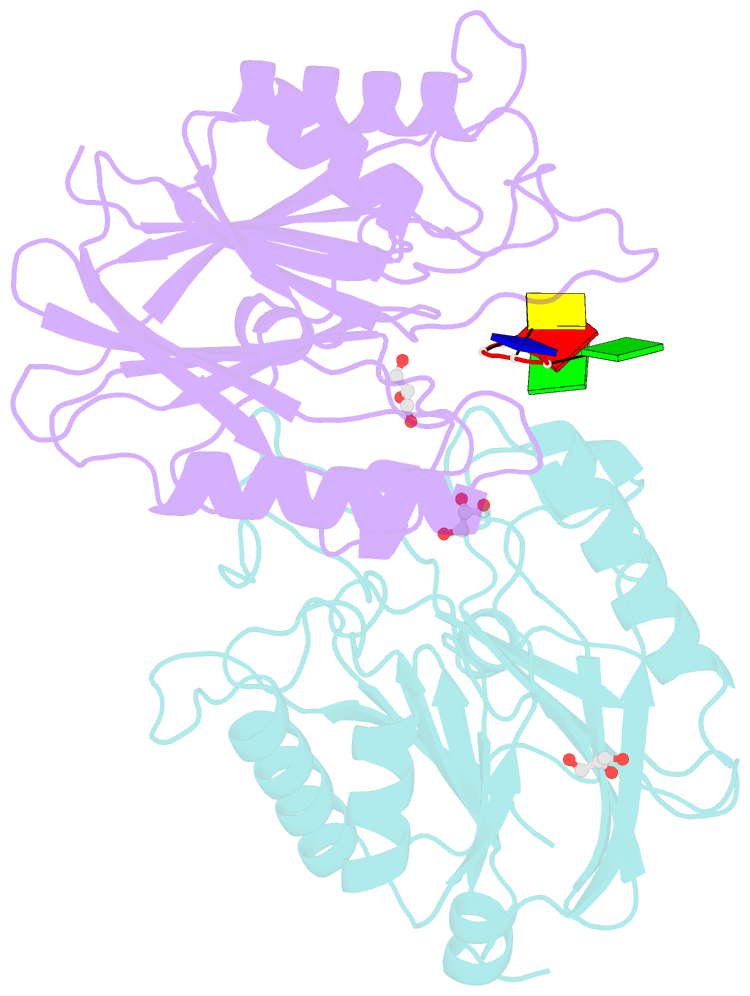
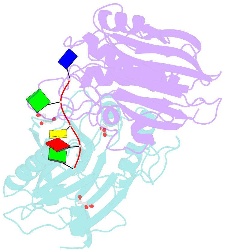
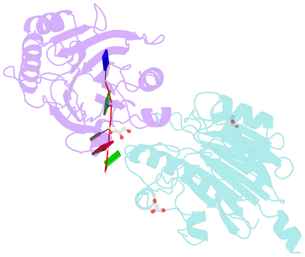
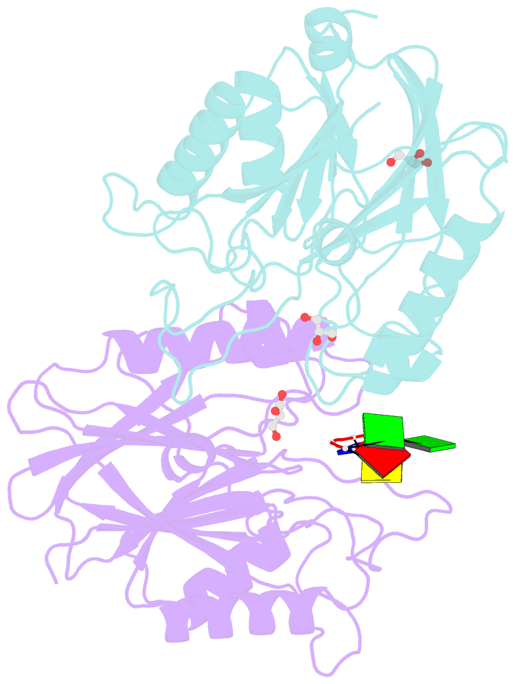
- Crystal structure of d. rerio tdp2 complexed with single strand DNA product
- Reference
- Shi K, Kurahashi K, Gao R, Tsutakawa SE, Tainer JA, Pommier Y, Aihara H (2012): "Structural basis for recognition of 5'-phosphotyrosine adducts by Tdp2." Nat.Struct.Mol.Biol., 19, 1372-1377. doi: 10.1038/nsmb.2423.
- Abstract
- The DNA-repair enzyme Tdp2 resolves 5'-phosphotyrosyl DNA adducts and mediates resistance to anticancer drugs that target covalent topoisomerase-DNA complexes. Tdp2 also participates in key signaling pathways during development and tumorigenesis and cleaves a protein-RNA linkage during picornavirus replication. The crystal structure of zebrafish Tdp2 bound to DNA reveals a deep, narrow basic groove that selectively accommodates the 5' end of single-stranded DNA in a stretched conformation. The crystal structure of the full-length Caenorhabditis elegans Tdp2 shows that this groove can also accommodate an acidic peptide stretch in vitro, with glutamate and aspartate side chains occupying the DNA backbone phosphate-binding sites. This extensive molecular mimicry suggests a potential mechanism for autoregulation and interaction of Tdp2 with phosphorylated proteins in signaling. Our study provides a framework to interrogate functions of Tdp2 and develop inhibitors for chemotherapeutic and antiviral applications.