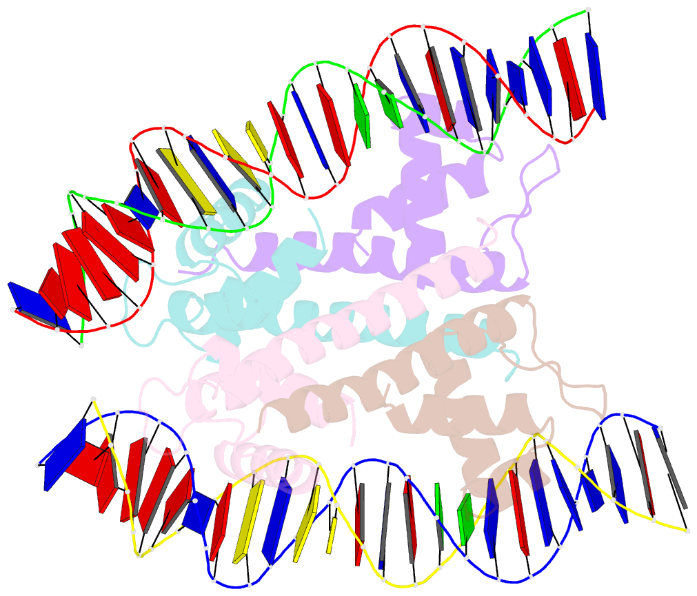
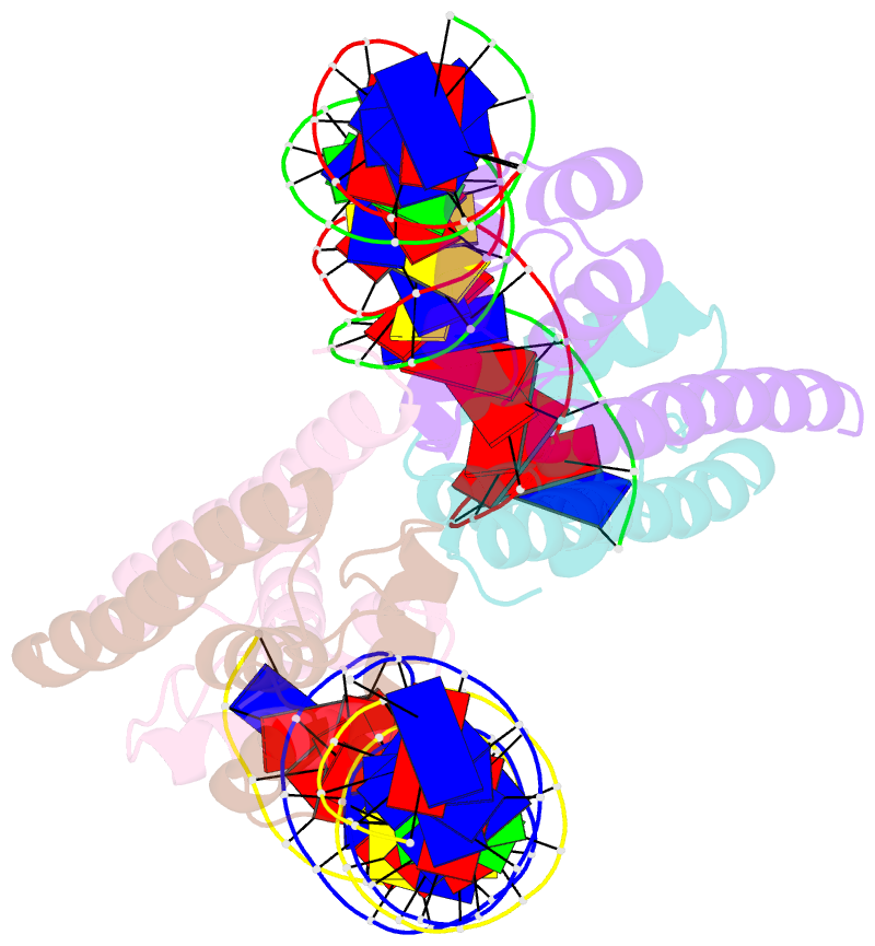
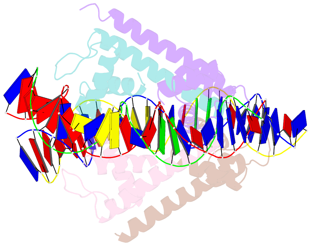
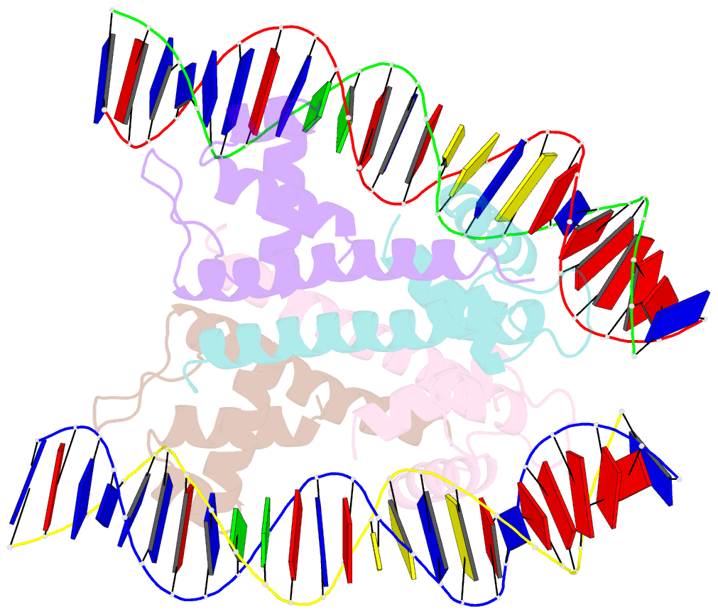
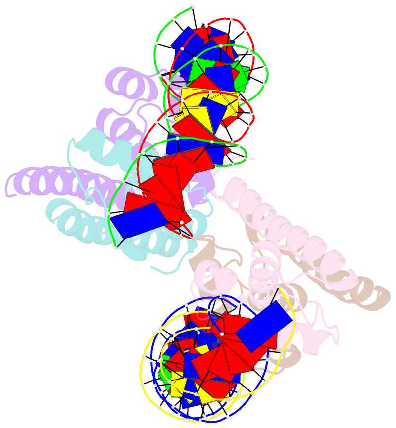
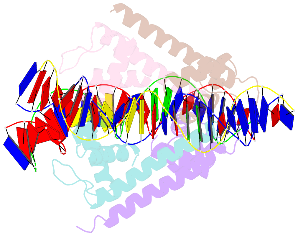
Summary information and primary citation
- PDB-id
- 4iht; SNAP-derived features in text and JSON formats;
DNAproDB
- Class
- transcription-DNA
- Method
- X-ray (3.0 Å)
- Summary
- Crystal structure of benm_dbd-bena site 1 DNA complex
- Reference
- Alanazi AM, Neidle EL, Momany C (2013): "The DNA-binding domain of BenM reveals the structural basis for the recognition of a T-N11-A sequence motif by LysR-type transcriptional regulators." Acta Crystallogr.,Sect.D, 69, 1995-2007. doi: 10.1107/S0907444913017320.
- Abstract
- LysR-type transcriptional regulators (LTTRs) play critical roles in metabolism and constitute the largest family of bacterial regulators. To understand protein-DNA interactions, atomic structures of the DNA-binding domain and linker-helix regions of a prototypical LTTR, BenM, were determined by X-ray crystallography. BenM structures with and without bound DNA reveal a set of highly conserved amino acids that interact directly with DNA bases. At the N-terminal end of the recognition helix (α3) of a winged-helix-turn-helix DNA-binding motif, several residues create hydrophobic pockets (Pro30, Pro31 and Ser33). These pockets interact with the methyl groups of two thymines in the DNA-recognition motif and its complementary strand, T-N11-A. This motif usually includes some dyad symmetry, as exemplified by a sequence that binds two subunits of a BenM tetramer (ATAC-N7-GTAT). Gln29 forms hydrogen bonds to adenine in the first position of the recognition half-site (ATAC). Another hydrophobic pocket defined by Ala28, Pro30 and Pro31 interacts with the methyl group of thymine, complementary to the base at the third position of the half-site. Arg34 interacts with the complementary base of the 3' position. Arg53, in the wing, provides AT-tract recognition in the minor groove. For DNA recognition, LTTRs use highly conserved interactions between amino acids and nucleotide bases as well as numerous less-conserved secondary interactions.