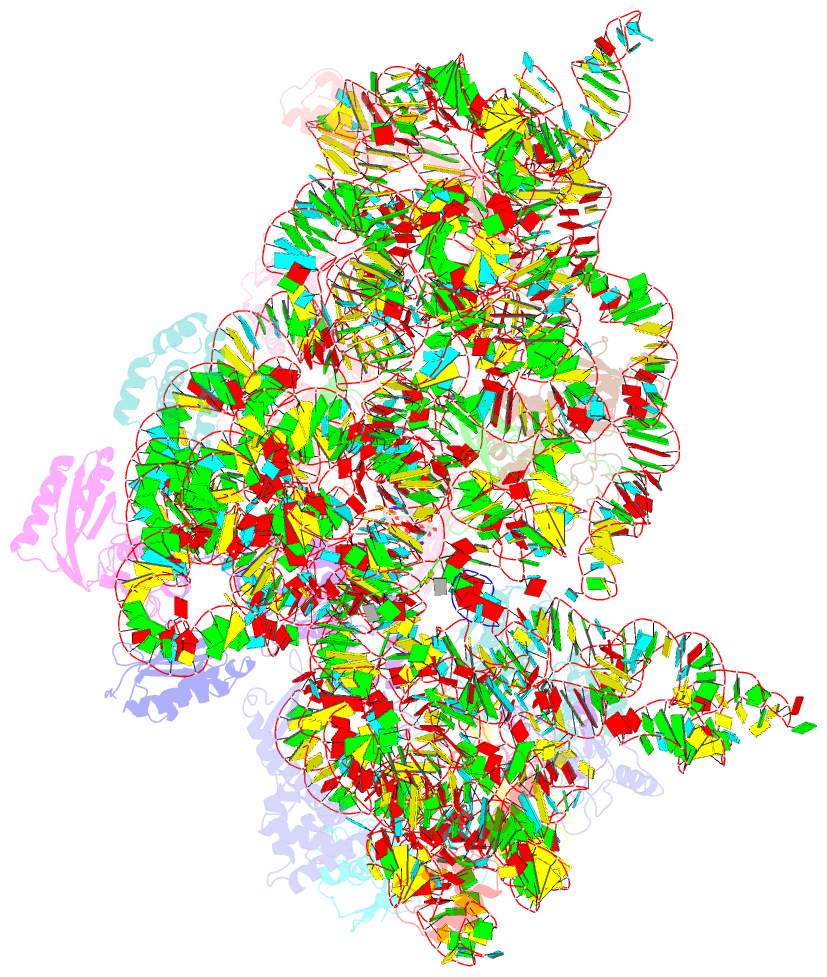
Summary information and primary citation
- PDB-id
- 4jya; SNAP-derived features in text and JSON formats;
DNAproDB
- Class
- ribosome
- Method
- X-ray (3.098 Å)
- Summary
- Crystal structures of pseudouridinilated stop codons with asls
- Reference
- Fernandez IS, Ng CL, Kelley AC, Wu G, Yu YT, Ramakrishnan V (2013): "Unusual base pairing during the decoding of a stop codon by the ribosome." Nature, 500, 107-110. doi: 10.1038/nature12302.
- Abstract
- During normal translation, the binding of a release factor to one of the three stop codons (UGA, UAA or UAG) results in the termination of protein synthesis. However, modification of the initial uridine to a pseudouridine (Ψ) allows efficient recognition and read-through of these stop codons by a transfer RNA (tRNA), although it requires the formation of two normally forbidden purine-purine base pairs. Here we determined the crystal structure at 3.1 Å resolution of the 30S ribosomal subunit in complex with the anticodon stem loop of tRNA(Ser) bound to the ΨAG stop codon in the A site. The ΨA base pair at the first position is accompanied by the formation of purine-purine base pairs at the second and third positions of the codon, which show an unusual Watson-Crick/Hoogsteen geometry. The structure shows a previously unsuspected ability of the ribosomal decoding centre to accommodate non-canonical base pairs.


