Summary information and primary citation
- PDB-id
- 4khp; SNAP-derived features in text and JSON formats;
DNAproDB
- Class
- ribosome-antibiotic
- Method
- X-ray (3.1 Å)
- Summary
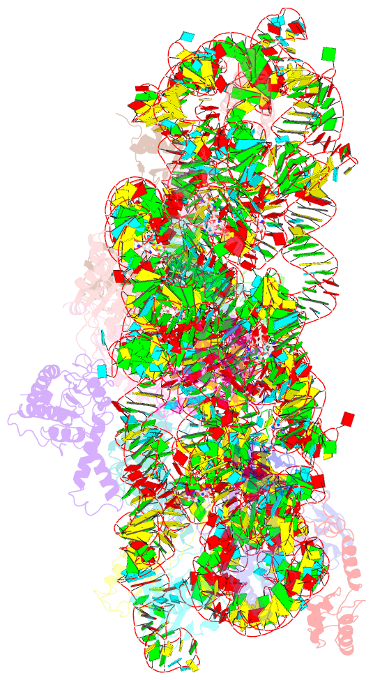
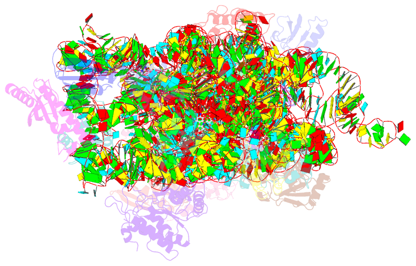
- Structure of the thermus thermophilus 30s ribosomal subunit in complex with de-6-msa-pactamycin
- Reference
- Tourigny DS, Fernandez IS, Kelley AC, Vakiti RR, Chattopadhyay AK, Dorich S, Hanessian S, Ramakrishnan V (2013): "Crystal Structure of a Bioactive Pactamycin Analog Bound to the 30S Ribosomal Subunit." J.Mol.Biol., 425, 3907-3910. doi: 10.1016/j.jmb.2013.05.004.
- Abstract
- Biosynthetically and chemically derived analogs of the antibiotic pactamycin and de-6-methylsalicylyl (MSA)-pactamycin have attracted recent interest as potential antiprotozoal and antitumor drugs. Here, we report a 3.1-Å crystal structure of de-6-MSA-pactamycin bound to its target site on the Thermus thermophilus 30S ribosomal subunit. Although de-6-MSA-pactamycin lacks the MSA moiety, it shares the same binding site as pactamycin and induces a displacement of nucleic acid template bound at the E-site of the 30S. The structure highlights unique interactions between this pactamycin analog and the ribosome, which paves the way for therapeutic development of related compounds.