Summary information and primary citation
- PDB-id
- 4oe1; SNAP-derived features in text and JSON formats;
DNAproDB
- Class
- RNA binding protein-RNA
- Method
- X-ray (2.8 Å)
- Summary
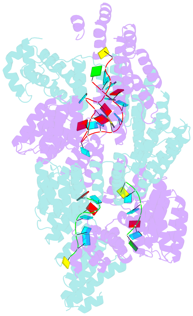
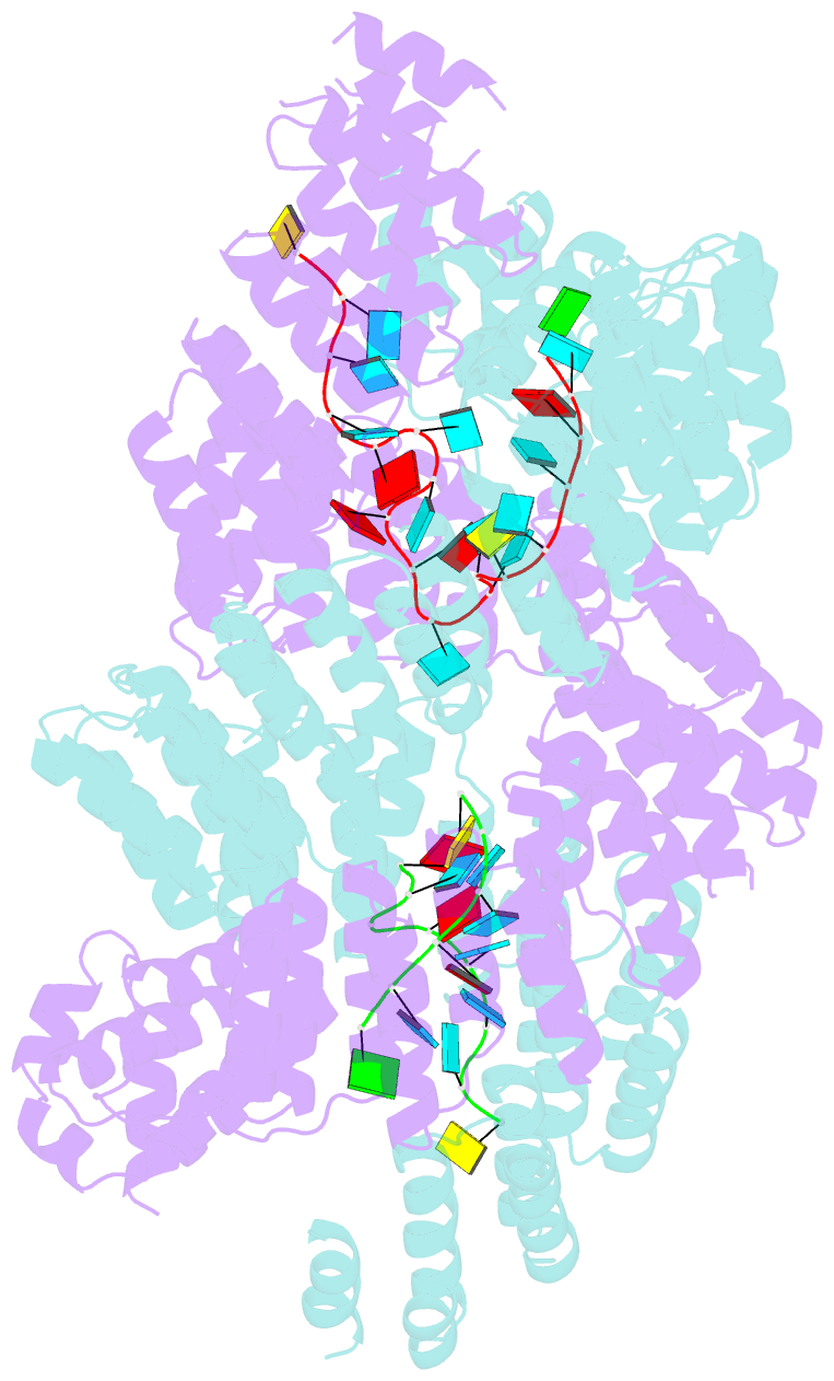
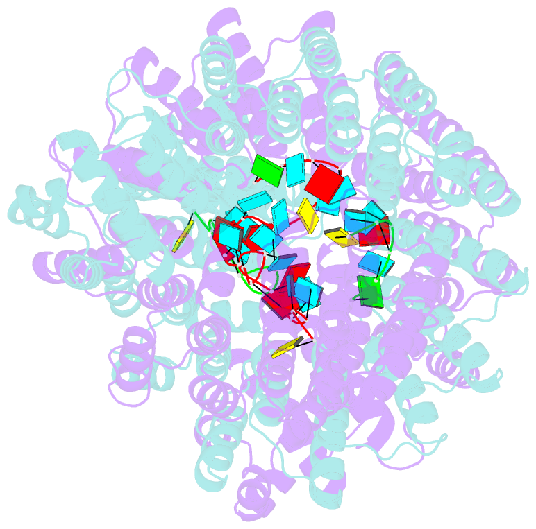
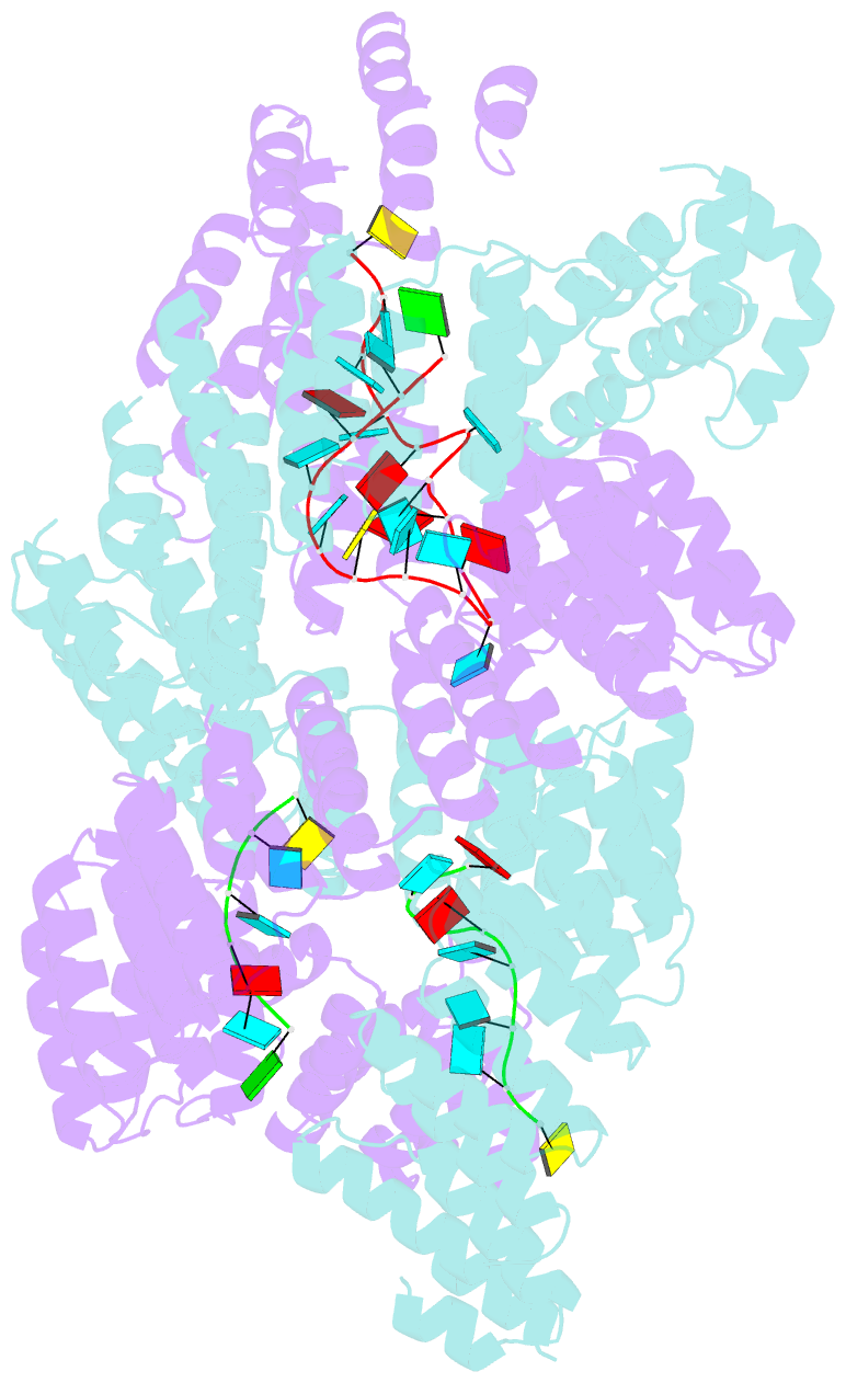
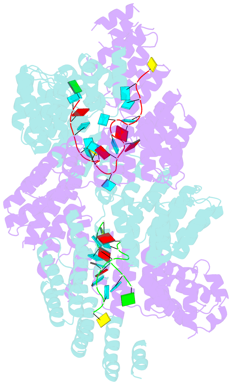
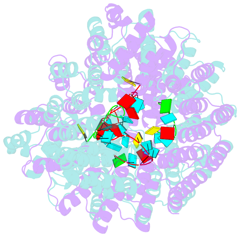
- Crystal structure of the pentatricopeptide repeat protein ppr10 (c256s-c430s-c449s) in complex with an 18-nt psaj RNA element
- Reference
- Li Q, Yan C, Xu H, Wang Z, Long J, Li W, Wu J, Yin P, Yan N (2014): "Examination of the dimerization states of the single-stranded RNA recognition protein pentatricopeptide repeat 10 (PPR10)." J.Biol.Chem., 289, 31503-31512. doi: 10.1074/jbc.M114.575472.
- Abstract
- Pentatricopeptide repeat (PPR) proteins, particularly abundant in plastids and mitochrondria of angiosperms, include a large number of sequence-specific RNA binding proteins that are involved in diverse aspects of organelle RNA metabolisms. PPR proteins contain multiple tandom repeats, and each repeat can specifically recognize a RNA base through residues 2, 5, and 35 in a modular fashion. The crystal structure of PPR10 from maize chloroplast exhibits dimeric existence both in the absence and presence of the 18-nucleotide psaJ RNA element. However, previous biochemical analysis suggested a monomeric shift of PPR10 upon RNA binding. In this report, we show that the amino-terminal segments of PPR10 determine the dimerization state of PPR10. A single amino acid alteration of cysteine to serine within repeat 10 of PPR10 further drives dimerization of PPR10. The biochemical elucidation of the determinants for PPR10 dimerization may provide an important foundation to understand the working mechanisms of PPR proteins underlying their diverse physiological functions.