Summary information and primary citation
- PDB-id
- 4pgq; SNAP-derived features in text and JSON formats;
DNAproDB
- Class
- transferase,lyase-DNA
- Method
- X-ray (2.3 Å)
- Summary
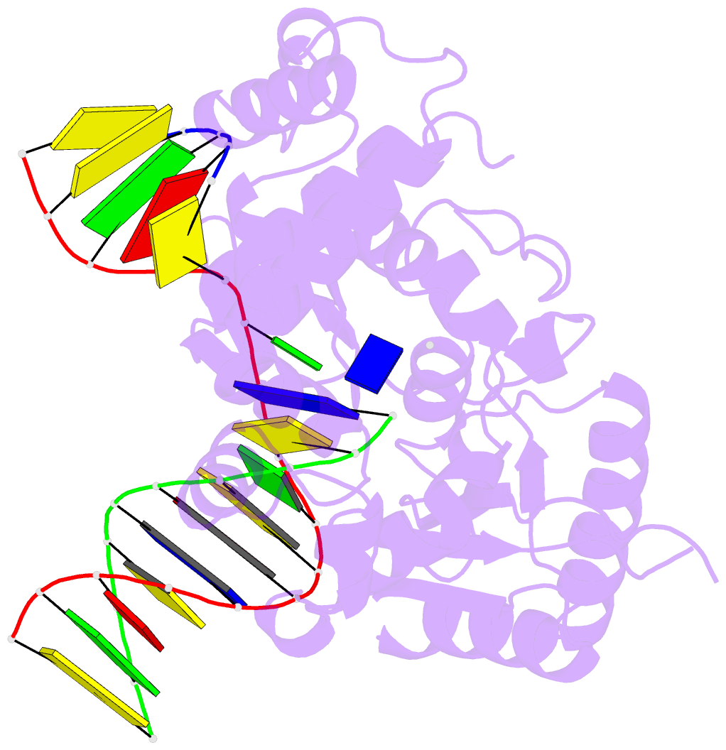
- Structure of human DNA polymerase beta complexed with g in the template base paired with incoming non-hydrolyzable ttp
- Reference
- Koag MC, Nam K, Lee S (2015): "The spontaneous replication error and the mismatch discrimination mechanisms of human DNA polymerase beta." Nucleic Acids Res., 42, 11233-11245. doi: 10.1093/nar/gku789.
- Abstract
- To provide molecular-level insights into the spontaneous replication error and the mismatch discrimination mechanisms of human DNA polymerase β (polβ), we report four crystal structures of polβ complexed with dG•dTTP and dA•dCTP mismatches in the presence of Mg2+ or Mn2+. The Mg(2+)-bound ground-state structures show that the dA•dCTP-Mg2+ complex adopts an 'intermediate' protein conformation while the dG•dTTP-Mg2+ complex adopts an open protein conformation. The Mn(2+)-bound 'pre-chemistry-state' structures show that the dA•dCTP-Mn2+ complex is structurally very similar to the dA•dCTP-Mg2+ complex, whereas the dG•dTTP-Mn2+ complex undergoes a large-scale conformational change to adopt a Watson-Crick-like dG•dTTP base pair and a closed protein conformation. These structural differences, together with our molecular dynamics simulation studies, suggest that polβ increases replication fidelity via a two-stage mismatch discrimination mechanism, where one is in the ground state and the other in the closed conformation state. In the closed conformation state, polβ appears to allow only a Watson-Crick-like conformation for purine•pyrimidine base pairs, thereby discriminating the mismatched base pairs based on their ability to form the Watson-Crick-like conformation. Overall, the present studies provide new insights into the spontaneous replication error and the replication fidelity mechanisms of polβ.


