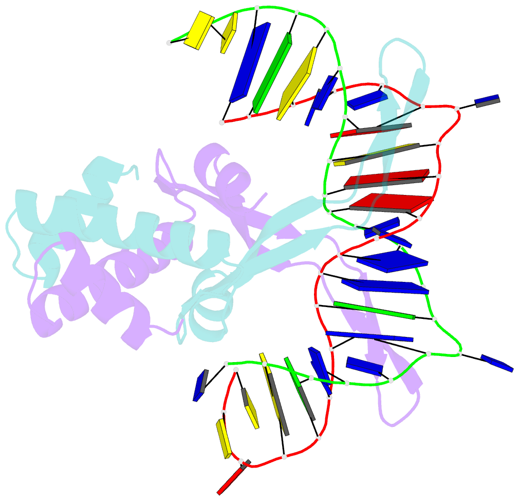
Summary information and primary citation
- PDB-id
- 4qju; SNAP-derived features in text and JSON formats;
DNAproDB
- Class
- DNA binding protein-DNA
- Method
- X-ray (2.16 Å)
- Summary
- Crystal structure of DNA-bound nucleoid associated protein, sav1473
- Reference
- Kim D-H, Im H, Jee J-G, Jang S-B, Yoon H-J, Kwon A-R, Kang S-M, Lee B-J (2014): "beta-Arm flexibility of HU from Staphylococcus aureus dictates the DNA-binding and recognition mechanism." Acta Crystallogr.,Sect.D, 70, 3273-3289. doi: 10.1107/S1399004714023931.
- Abstract
- HU, one of the major nucleoid-associated proteins, interacts with the minor groove of DNA in a nonspecific manner to induce DNA bending or to stabilize bent DNA. In this study, crystal structures are reported for both free HU from Staphylococcus aureus Mu50 (SHU) and SHU bound to 21-mer dsDNA. The structures, in combination with electrophoretic mobility shift assays (EMSAs), isothermal titration calorimetry (ITC) measurements and molecular-dynamics (MD) simulations, elucidate the overall and residue-specific changes in SHU upon recognizing and binding to DNA. Firstly, structural comparison showed the flexible nature of the β-sheets of the DNA-binding domain and that the β-arms bend inwards upon complex formation, whereas the other portions are nearly unaltered. Secondly, it was found that the disruption and formation of salt bridges accompanies DNA binding. Thirdly, residue-specific free-energy analyses using the MM-PBSA method with MD simulation data suggested that the successive basic residues in the β-arms play a central role in recognizing and binding to DNA, which was confirmed by the EMSA and ITC analyses. Moreover, residue Arg55 resides in the hinge region of the flexible β-arms, exhibiting a remarkable role in their flexible nature. Fourthly, EMSAs with various DNAs revealed that SHU prefers deformable DNA. Taken together, these data suggest residue-specific roles in local shape and base readouts, which are primarily mediated by the flexible β-arms consisting of residues 50-80.


